Eukaryotic cells and cell organelles
NCERT Class 11 Biology Chapter 8 Section 8.5
Quick answer
Typical eukaryotic cells have a nucleus and membrane-bound organelles, with ER, Golgi, lysosomes and vacuoles forming NCERT’s endomembrane system. Mitochondria and chloroplasts have double membranes, DNA and ribosomes; plant cells typically also have a wall, plastids and a large vacuole. Specialised cells can lose components, as mature mammalian red cells lose their nucleus.
With your textbook
NCERT Biology (Class 11), Chapter 8 “Cell: The Unit of Life”, Section 8.5 “Eukaryotic Cells”, pages 91–102.
Open the official NCERT chapter (PDF, 7.0 MB, ncert.nic.in) (opens in a new tab)
Checked against the 2026-27 reprint on .
Eukaryotic cells
Eukaryotic cells
The eukaryotes include all the protists, plants, animals and fungi.
NCERT Biology (Class 11), Section 8.5, page 91
- The cytoplasm is extensively compartmentalised by membrane-bound organelles (page 91).
- The cell has an organised nucleus with a nuclear envelope, and its genetic material is organised into chromosomes (page 91).
- Eukaryotic cells also have a variety of complex locomotory and cytoskeletal structures (page 91).
In plain words: a eukaryotic cell is divided into rooms. Most of its DNA has its own room, the nucleus, and other jobs are done in separate membrane-bound organelles: proteins made by ribosomes on the rough ER are modified and packaged in the Golgi apparatus, energy is released in mitochondria, and in plants food is made in chloroplasts. This page takes the organelles one by one, in NCERT’s order.
Plant and animal cells
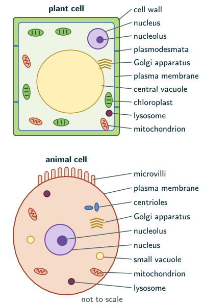
All eukaryotic cells are not identical (page 91). The table compares typical plant and animal cells; specialised cells can differ, as some mature cells even lack a nucleus (page 100):
| Plant cell | Animal cell |
|---|---|
| cell wall | no cell wall |
| plastids | no plastids |
| large central vacuole | no large central vacuole |
| centrioles absent in almost all | centrioles present |
Cell membrane
The detailed structure of the membrane was studied only in the 1950s, with the electron microscope; before that, chemical studies, especially on human red blood cells, suggested its structure (page 93).
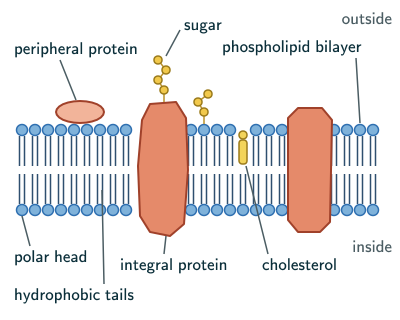
- The membrane is mainly lipids and proteins. The major lipids are phospholipids, arranged in a bilayer with the polar heads towards the outer sides and the hydrophobic tails towards the inner part, so the nonpolar tails are protected from the watery surroundings. The membrane also contains cholesterol (page 93).
- Membranes also have protein and carbohydrate. The ratio of protein to lipid varies between cell types: the membrane of the human erythrocyte has approximately 52 per cent protein and 40 per cent lipids (page 93).
- By ease of extraction, membrane proteins are integral (partially or totally buried in the membrane) or peripheral (on its surface) (page 93).
Fluid mosaic model. Singer and Nicolson (1972) proposed the widely accepted fluid mosaic model: the quasi-fluid nature of the lipid lets proteins move sideways within the bilayer. This ability to move is measured as the membrane’s fluidity, which matters for cell growth, formation of intercellular junctions, secretion, endocytosis and cell division (page 94).
Transport across the membrane. The membrane is selectively permeable (page 94):
- Passive transport needs no energy. Neutral solutes move by simple diffusion along the concentration gradient, from higher to lower concentration. Water also moves from higher to lower concentration of water, that is, from a dilute solution towards a more concentrated one; the movement of water by diffusion is osmosis.
- Polar molecules cannot pass through the nonpolar lipid bilayer, so they need a carrier protein of the membrane to help them across.
- Active transport moves a few ions or molecules against their concentration gradient, from lower to higher concentration. It is energy dependent and uses ATP, as in the Na/K pump (page 94).
Reading note (page 94): Beyond the NCERT text: polarity is a matter of degree. Most polar molecules cross the bilayer poorly and charged ones worse still, but water, though polar, crosses it fairly easily, and water channels in the membrane, the aquaporins, let it flow faster still (Wikipedia, “Lipid bilayer” and “Aquaporin”, facts in our words). Active transport that uses ATP directly is primary active transport; secondary active transport is driven instead by an ion moving down its electrochemical gradient, which can differ from its concentration gradient (Wikipedia, “Active transport”, facts in our words). In an exam, give NCERT’s account.
Movement of water by diffusion is called osmosis.
NCERT Biology (Class 11), Section 8.5.1, page 94
Cell wall
A non-living rigid structure, the cell wall, covers the plasma membrane of fungi and plants (page 94).
- It gives the cell its shape, protects it from mechanical damage and infection, helps in cell-to-cell interaction and is a barrier to undesirable macromolecules.
- Algae: cellulose, galactans, mannans and minerals such as calcium carbonate. Other plants: cellulose, hemicellulose, pectins and proteins. Fungi: chitin and polysaccharides (Kingdom Fungi, NCERT Section 2.3, page 16).
- The primary wall of a young plant cell can grow; this ability diminishes as the cell matures, and the secondary wall forms on its inner side, towards the membrane (page 94). Beyond the NCERT text: many plant cells, not all, lay down a secondary wall, and only when they have finished enlarging, with the primary wall already in place (Wikipedia, “Secondary cell wall”, facts in our words).
- The middle lamella, mainly calcium pectate, glues neighbouring cells together. The wall and middle lamellae may be crossed by plasmodesmata, which connect the cytoplasm of neighbouring cells (page 94).
The endomembrane system
The ER, Golgi complex, lysosomes and vacuoles are considered together as the endomembrane system, because their functions are coordinated. Mitochondria, chloroplasts and peroxisomes are not part of it, as their functions are not coordinated with these (page 95).
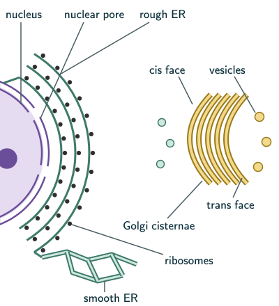
Endoplasmic reticulum (ER). A network of tiny tubular structures scattered in the cytoplasm. It divides the space inside the cell into a luminal compartment (inside the ER) and an extra-luminal compartment (the cytoplasm) (page 95).
- Rough ER (RER) bears ribosomes on its outer surface. It is common in cells actively making and secreting proteins, and is extensive and continuous with the outer membrane of the nucleus.
- Smooth ER (SER) has no ribosomes. It is the major site of lipid synthesis; in animal cells, lipid-like steroidal hormones are made in the SER (page 95).
Golgi apparatus. Camillo Golgi (1898) first saw densely stained reticular structures near the nucleus, later named Golgi bodies (page 95).
- It is a stack of flat, disc-shaped sacs, the cisternae, 0.5 µm to 1.0 µm in diameter, arranged near the nucleus with a convex cis (forming) face and a concave trans (maturing) face (pages 95–96).
- Its main job is packaging materials for targets inside the cell or for secretion. Vesicles from the ER fuse with the cis face and move towards the trans face, which is why the Golgi stays close to the ER. Many proteins made on the ER are modified in the cisternae before release from the trans face (page 96).
Golgi apparatus is the important site of formation of glycoproteins and glycolipids.
NCERT Biology (Class 11), Section 8.5.3, page 96
Lysosomes. Membrane-bound vesicles formed by packaging in the Golgi apparatus. They are very rich in almost all types of hydrolytic enzymes (lipases, proteases, carbohydrases), most active at acidic pH, which can digest carbohydrates, proteins, lipids and nucleic acids (page 96).
Vacuoles. A membrane-bound space in the cytoplasm containing water, sap, excretory products and other materials not useful to the cell (page 96).
- It is bound by a single membrane, the tonoplast. In plant cells, vacuoles can occupy up to 90 per cent of the cell’s volume.
- In plants, the tonoplast moves ions and other materials into the vacuole against their concentration gradients, so their concentration is much higher in the vacuole than in the cytoplasm.
- In Amoeba, the contractile vacuole is important for osmoregulation and excretion; in many cells, as in protists, food vacuoles form by engulfing food particles (page 96).
Mitochondria
Unless specially stained, mitochondria are not easily seen under the microscope. Their number per cell varies with the cell’s physiological activity, and their shape and size vary too (page 96).
- Typically sausage-shaped or cylindrical, with a diameter of 0.2–1.0 µm (average 0.5 µm) and a length of 1.0–4.1 µm (page 96).
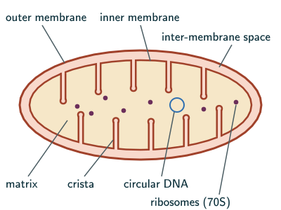
- Double membrane-bound. The outer membrane is the continuous limiting boundary. The inner membrane forms infoldings, the cristae, towards the matrix, which increase the surface area. The two membranes divide the lumen into an outer compartment and an inner compartment filled with the dense matrix (page 97).
- The matrix has a single circular DNA molecule, a few RNA molecules, 70S ribosomes and the components needed to make proteins. Mitochondria divide by fission (page 97). Beyond the NCERT text: mitochondrial ribosomes differ between groups; those of mammals are smaller, 55S (39S and 28S subunits) (Wikipedia, “Mitochondrial ribosome”, facts in our words). In an NCERT-based answer, give 70S.
Mitochondria are the sites of aerobic respiration.
NCERT Biology (Class 11), Section 8.5.4, page 97
They produce cellular energy as ATP, which is why they are called the “power houses” of the cell (page 97).
Plastids
Plastids are found in all plant cells and in euglenoids (NCERT spells it “euglenoides”). They are large and easy to see, and their pigments give plant parts their colours (page 97).
- Chloroplasts: chlorophyll and carotenoid pigments, which trap the light energy essential for photosynthesis.
- Chromoplasts: fat-soluble carotenoid pigments such as carotene and xanthophylls, giving yellow, orange or red colours.
- Leucoplasts: colourless plastids that store nutrients: amyloplasts store carbohydrates (starch), as in potato; elaioplasts store oils and fats; aleuroplasts store proteins (pages 97–98).
NCERT’s chapter Summary calls plastids “pigment containing organelles found in plant cells only” (page 102). The section itself says they occur in euglenoids too, and that leucoplasts are colourless (page 97); go by the section.
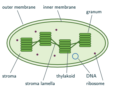
The chloroplast (page 98):
- Most chloroplasts of green plants are in the mesophyll cells of leaves. They are lens-shaped, oval, spherical, discoid or even ribbon-like, 5–10 µm long and 2–4 µm wide; their number ranges from 1 per cell in Chlamydomonas, a green alga, to 20–40 per mesophyll cell.
- Double membrane-bound; the inner membrane is relatively less permeable. The space inside it is the stroma.
- In the stroma, flattened membranous sacs, the thylakoids, are stacked like piles of coins as grana (singular: granum). Flat membranous tubules, the stroma lamellae, connect the thylakoids of different grana. The space inside a thylakoid is its lumen.
- Chlorophyll is in the thylakoids. The stroma has enzymes for making carbohydrates and proteins, small double-stranded circular DNA and ribosomes; chloroplast ribosomes are 70S, smaller than the cytoplasmic 80S ribosomes.
- NCERT’s Summary adds that the grana are the site of the light reactions and the stroma of the dark reactions of photosynthesis (page 102).
NCERT’s sentence on grana adds “or the intergranal thylakoids” after “grana” (page 98), which reads as if grana had that second name. The thylakoids between grana are the stroma lamellae, which NCERT describes in the next sentence; a granum is a stack of thylakoids.
Ribosomes
Ribosomes are the granular structures first observed under the electron microscope as dense particles by George Palade (1953).
NCERT Biology (Class 11), Section 8.5.6, page 98
- Ribosomes are the site of protein synthesis (page 91). In eukaryotic cells they occur in the cytoplasm, on the rough ER and the outer nuclear membrane, and inside mitochondria and chloroplasts (pages 88 and 100).
- Ribosomes are made of RNA and proteins and are not surrounded by any membrane (page 98).
- Eukaryotic ribosomes are 80S, with subunits of 60S and 40S; prokaryotic ribosomes are 70S, with subunits of 50S and 30S (page 98).
- S (Svedberg unit) is the sedimentation coefficient, indirectly a measure of density and size (page 98). S values of the subunits do not add up to the whole: 60S and 40S make an 80S ribosome (our reasoning, from NCERT page 98).
Cytoskeleton
The cytoskeleton is the network of filamentous protein structures in the cytoplasm: microtubules, microfilaments and intermediate filaments. It gives mechanical support, helps in motility and maintains the shape of the cell (page 98).
Cilia and flagella
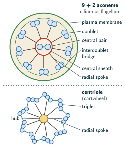
- Cilia and flagella are hair-like outgrowths of the cell membrane. Cilia are small and work like oars, moving either the cell or the fluid around it (see ciliary movement); flagella are longer and move the cell. Bacterial flagella are structurally different from eukaryotic flagella (page 99).
- Both are covered with plasma membrane. Their core, the axoneme, has microtubules running along its length: usually nine doublets of peripheral microtubules arranged radially and a pair of central microtubules, the 9+2 array (page 99).
- The central tubules are joined by bridges and enclosed by a central sheath, which is connected to one tubule of each peripheral doublet by a radial spoke, so there are nine radial spokes. The peripheral doublets are interconnected by linkers. Both cilia and flagella arise from centriole-like basal bodies (page 99).
Centrosome and centrioles
- The centrosome usually contains two cylindrical centrioles, surrounded by amorphous pericentriolar materials. The two centrioles lie perpendicular to each other, each organised like a cartwheel (page 99).
- Each centriole has nine evenly spaced peripheral fibrils of tubulin protein; each fibril is a triplet, and adjacent triplets are linked. The proteinaceous central part of its proximal region, the hub, is joined to the triplets by radial spokes of protein (page 100).
- Centrioles form the basal bodies of cilia and flagella, and in animal cells the spindle fibres that give rise to the spindle apparatus during cell division (page 100). Beyond the NCERT text: the microtubules are started (nucleated) by proteins in the pericentriolar material round the two centrioles, and these centrosome microtubules build the mitotic spindle (Wikipedia, “Centrosome”, facts in our words). In an NCERT-based answer, give NCERT’s line.
Nucleus
Nucleus as a cell organelle was first described by Robert Brown as early as 1831.
NCERT Biology (Class 11), Section 8.5.10, page 100
- Flemming later named the material of the nucleus that stains with basic dyes chromatin (page 100).
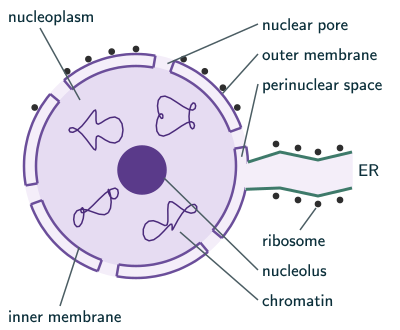
- The interphase nucleus (of a cell that is not dividing) has extended nucleoprotein fibres, the chromatin, a nuclear matrix and one or more spherical nucleoli (page 100).
- The nuclear envelope has two parallel membranes with a space of 10 to 50 nm between them, the perinuclear space. It is a barrier between the contents of the nucleus and the cytoplasm. The outer membrane usually remains continuous with the ER and bears ribosomes (page 100).
- Nuclear pores, formed where the two membranes fuse, interrupt the envelope at many places. RNA and protein molecules pass through them in both directions between the nucleus and the cytoplasm (page 100).
- Normally a cell has one nucleus, but the number varies (page 100): the coenocytic hyphae of some fungi are continuous tubes of multinucleate cytoplasm (NCERT Section 2.3, page 16), and the liquid endosperm of coconut is a multinucleate syncytium (NCERT Section 10.2, page 124). Some mature cells lack a nucleus, such as the erythrocytes of many mammals and the sieve tube cells of vascular plants (page 100). Beyond the NCERT text: the red blood cells of mammals lack a nucleus when mature, unlike those of other vertebrates (Wikipedia, “Red blood cell”, facts in our words).
- The nucleoplasm contains the nucleolus and the chromatin. The nucleolus is not membrane-bound, so its content is continuous with the nucleoplasm; it is a site of active ribosomal RNA synthesis, and cells making much protein have larger and more numerous nucleoli (page 100).
Chromatin and chromosomes (pages 101–102):
- In the interphase nucleus, chromatin is a loose, indistinct network; during cell division, structured chromosomes appear in place of the nucleus.
- Chromatin contains DNA, basic proteins called histones, some non-histone proteins and RNA. A single human cell has about two metres of DNA, distributed among its 46 chromosomes (23 pairs) (page 101). These 46, in 23 pairs, are the diploid (2n) set: a gamete, which is haploid, has 23, and in S phase the DNA doubles (2C to 4C) while the number of chromosomes stays 46 (reasoned from NCERT Chapter 10, pages 121 and 125).
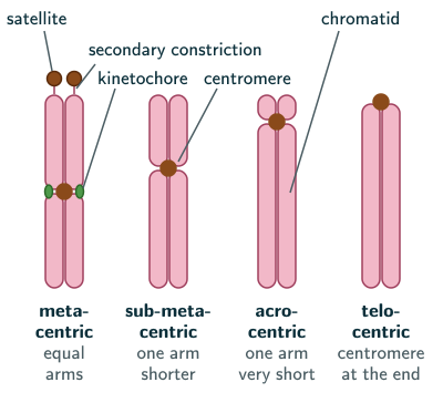
- Every chromosome has a primary constriction, the centromere, which holds the two chromatids; disc-shaped kinetochores lie on its sides (page 101).
- By the position of the centromere, chromosomes are of four types: metacentric (middle, two equal arms), sub-metacentric (slightly away from the middle, one shorter and one longer arm), acrocentric (close to the end, one extremely short and one very long arm) and telocentric (terminal).
- A few chromosomes have non-staining secondary constrictions at a constant place, giving the appearance of a small fragment, the satellite (page 102); the satellite stays joined to the rest of the chromosome (our reading of NCERT’s “appearance”).
NCERT’s Figure 8.13 does not name its four chromosomes, and it labels a short arm even on the last one. A telocentric chromosome, with its centromere at the very end, has only one arm.
Microbodies
Many membrane bound minute vesicles called microbodies that contain various enzymes, are present in both plant and animal cells.
NCERT Biology (Class 11), Section 8.5.11, page 102
Solved examples
Example 1: Match the structures (NCERT Exercise 3)
Match (a) cristae, (b) cisternae, (c) thylakoids with (i) flat membranous sacs in stroma, (ii) infoldings in mitochondria, (iii) disc-shaped sacs in Golgi apparatus.
Answer: (a) ii, (b) iii, (c) i (NCERT pages 95–98).
Example 2: Neutral and polar molecules (NCERT Exercise 6)
How do neutral solutes cross the plasma membrane? Can polar molecules cross the same way?
Answer: neutral solutes move by simple diffusion along the concentration gradient, from higher to lower concentration, with no energy. Polar molecules cannot pass through the nonpolar lipid bilayer; they need a carrier protein of the membrane. Some ions and molecules are moved against the gradient by active transport, using ATP, as in the Na/K pump (NCERT page 94). Beyond the NCERT text: water is the notable polar exception; it crosses the bilayer fairly easily, and faster through aquaporins (see the reading note above).
Example 3: Two double membrane-bound organelles (NCERT Exercise 7)
Name two double membrane-bound organelles and state their functions.
Answer: mitochondria and chloroplasts. A mitochondrion is sausage-shaped or cylindrical, 0.2–1.0 µm wide and 1.0–4.1 µm long; its outer membrane is the continuous boundary and its inner membrane folds into cristae towards the matrix, which holds a single circular DNA, a few RNA molecules and 70S ribosomes (55S in mammals, beyond the NCERT text: see the mitochondria section); it divides by fission and is the site of aerobic respiration, making ATP. A chloroplast is lens-shaped to ribbon-like, 5–10 µm long and 2–4 µm wide; its less permeable inner membrane encloses the stroma, where thylakoids are stacked as grana joined by stroma lamellae; chlorophyll is in the thylakoids, and the stroma has enzymes, circular DNA and 70S ribosomes; it traps light energy for photosynthesis (NCERT pages 96–98). Draw them as in the mitochondrion and chloroplast figures above. The nucleus is also bounded by two membranes, its envelope (page 100).
Example 4: Lysosomes and vacuoles (NCERT Exercise 12)
Both lysosomes and vacuoles belong to the endomembrane system. How do their functions differ?
Answer: lysosomes are packed with hydrolytic enzymes, active at acidic pH, that digest carbohydrates, proteins, lipids and nucleic acids. Vacuoles store water, sap, excretory products and other materials not useful to the cell; in plants the tonoplast concentrates ions in them, and in Amoeba the contractile vacuole works in osmoregulation and excretion (NCERT page 96).
Example 5: DNA per chromosome
A human cell has about 2 m of DNA in 46 chromosomes. About how long is the DNA in one chromosome, on average?
Answer: 2 m ÷ 46 ≈ 0.043 m, about 4.3 cm (DNA length and chromosome number from NCERT page 101).
Example 6: Counting microtubules
How many microtubules are there in the axoneme of a cilium, and in a centriole?
Answer: axoneme: nine doublets (9 × 2 = 18) plus the central pair (2), 20 in all. Centriole: nine triplets, 9 × 3 = 27, with no central pair (NCERT pages 99–100).
Common mistakes to avoid
Common mistake
Swapping the cis and trans faces
The cis face is the forming face, convex, where vesicles from the ER arrive; the trans face is the maturing face, concave, from which material leaves (NCERT pages 95–96).
Common mistake
Giving mitochondria or chloroplasts 80S ribosomes
Their ribosomes are 70S; the 80S ribosomes are in the eukaryotic cytoplasm (NCERT pages 97–98). Beyond the NCERT text: mammalian mitochondrial ribosomes are 55S (see the mitochondria section); in an NCERT-based answer, give 70S.
Common mistake
Calling the nucleolus membrane-bound
The nucleolus has no membrane; its content is continuous with the nucleoplasm (NCERT page 100).
Common mistake
Putting mitochondria in the endomembrane system
The endomembrane system is the ER, Golgi complex, lysosomes and vacuoles; mitochondria, chloroplasts and peroxisomes are not part of it (NCERT page 95).
Quick revision summary
- Eukaryotes: protists, plants, animals, fungi; membrane-bound organelles, an organised nucleus, chromosomes. Plant cells: wall, plastids, large central vacuole; animal cells: centrioles.
- Membrane: phospholipid bilayer, cholesterol, proteins (integral, peripheral) and carbohydrate; fluid mosaic model (Singer and Nicolson, 1972); diffusion, osmosis, carrier proteins, active transport (Na/K pump).
- Cell wall: algae (cellulose, galactans, mannans, calcium carbonate), other plants (cellulose, hemicellulose, pectins, proteins); primary and secondary walls; middle lamella of calcium pectate; plasmodesmata.
- Endomembrane system: RER (proteins) and SER (lipids, steroid hormones); Golgi (cis and trans faces, packaging, glycoproteins, glycolipids); lysosomes (acid hydrolases); vacuoles (tonoplast, up to 90 per cent of a plant cell).
- Mitochondria: double membrane, cristae, matrix with circular DNA and 70S ribosomes (55S in mammals, beyond the NCERT text), aerobic respiration, ATP, fission. Plastids: chloroplasts, chromoplasts, leucoplasts (amyloplasts, elaioplasts, aleuroplasts); chloroplast stroma, grana, stroma lamellae.
- Ribosomes 80S and 70S; cytoskeleton; cilia and flagella (9+2); centrioles (nine triplets, cartwheel), basal bodies and spindle.
- By membranes: none, ribosomes, the centrosome (NCERT page 88) and the nucleolus (page 100); one, lysosomes (page 102) and vacuoles (the tonoplast, page 96); two, the nucleus (its envelope), mitochondria and chloroplasts (pages 97–100).
- Nucleus: envelope with perinuclear space and pores, nucleolus (rRNA), chromatin; chromosomes with centromere and kinetochores, four types by centromere position; satellite; microbodies.
Formula sheet
- endomembrane system: ER, Golgi complex, lysosomes, vacuoles
- ribosomes: 80S (60S, 40S) cytoplasm; 70S (50S, 30S) prokaryotes, mitochondria, chloroplasts
- mitochondrion: 0.2–1.0 µm wide (average 0.5), 1.0–4.1 µm long
- chloroplast: 5–10 µm long, 2–4 µm wide
- axoneme: 9 + 2 (nine doublets, a central pair)
- centriole: nine triplets, cartwheel
- perinuclear space: 10–50 nm
- chromosome types: metacentric, sub-metacentric, acrocentric, telocentric
Questions students ask
What is the difference between a plant cell and an animal cell?
Plant and animal cells are both eukaryotic, but NCERT says plant cells possess cell walls, plastids and a large central vacuole, which are absent in animal cells, while animal cells have centrioles, which are absent in almost all plant cells (page 91). NCERT’s Figure 8.3 also labels plasmodesmata and the middle lamella in the plant cell and microvilli in the animal cell. Both have a plasma membrane, a nucleus with a nucleolus, rough and smooth ER, a Golgi apparatus, mitochondria, ribosomes, lysosomes and peroxisomes.
What are the main cell organelles and their functions?
The main cell organelles and their roles, in NCERT’s account: the nucleus controls the activities of the organelles and plays a major role in heredity; ribosomes are the site of protein synthesis; the rough ER works in protein synthesis and secretion, the smooth ER in lipid synthesis; the Golgi apparatus packages materials and forms glycoproteins and glycolipids; lysosomes digest all types of macromolecules; and vacuoles hold water, sap and excretory products. Mitochondria are the sites of aerobic respiration, producing ATP, and chloroplasts trap light energy for photosynthesis. Centrioles form basal bodies of cilia and flagella; centrosomes help organise the spindle in many animal cells.
What is the fluid mosaic model, and why is it called so?
The fluid mosaic model, proposed by Singer and Nicolson (1972), is the widely accepted model of the cell membrane: a phospholipid bilayer, polar heads outwards and hydrophobic tails inwards, with integral proteins partly or totally buried in it and peripheral proteins on its surface. It is called fluid because the quasi-fluid nature of the lipid enables lateral movement of proteins within the bilayer, an ability measured as the membrane’s fluidity, and mosaic because, beyond the NCERT text, the scattered proteins look like the pieces of a mosaic. NCERT links this fluidity to cell growth, intercellular junctions, secretion, endocytosis and cell division.
What is the difference between 70S and 80S ribosomes?
80S ribosomes are the eukaryotic ribosomes of the cytoplasm, with subunits of 60S and 40S, while 70S ribosomes, with subunits of 50S and 30S, occur in prokaryotes and, in NCERT’s account, inside mitochondria and chloroplasts (NCERT pages 97–98). So, in that account, a typical eukaryotic cell has both kinds: 80S in its cytoplasm and on the rough ER, and 70S in its mitochondria and, in plants, its chloroplasts. S is the Svedberg unit, the sedimentation coefficient, which NCERT calls indirectly a measure of density and size; it is not a mass, so subunit S values do not add up to the whole. All these ribosomes are made of RNA and proteins, have no membrane and are the sites of protein synthesis.
What is the difference between the cell wall and the cell membrane?
The cell membrane, or plasma membrane, is a selectively permeable layer of lipids and proteins that bounds every cell, while the cell wall is a non-living rigid structure that NCERT says forms an outer covering for the plasma membrane of fungi and plants (page 94). The membrane, a phospholipid bilayer with integral and peripheral proteins, controls what enters and leaves the cell; the wall gives the cell its shape, protects it from mechanical damage and infection and is a barrier to undesirable macromolecules. Animal cells have only a membrane; most plant walls consist of cellulose, hemicellulose, pectins and proteins, and most prokaryotes have one; mycoplasmas are NCERT’s wall-less example.
What is the difference between rough and smooth ER?
The rough endoplasmic reticulum (RER) bears ribosomes on its outer surface, while the smooth endoplasmic reticulum (SER) has none and so appears smooth. RER is frequently observed in cells actively involved in protein synthesis and secretion and is extensive and continuous with the outer membrane of the nucleus, while SER is the major site for synthesis of lipid, and in animal cells lipid-like steroidal hormones are synthesised in it (NCERT page 95). Both belong to the endomembrane system, with the Golgi complex, lysosomes and vacuoles.
Why are mitochondria and chloroplasts called semi-autonomous?
Mitochondria and chloroplasts are called semi-autonomous organelles because each has its own DNA and ribosomes and can make some of its own proteins: NCERT says the mitochondrial matrix has circular DNA, RNA and ribosomes (70S in NCERT’s simplified account) and the components required for the synthesis of proteins, and the chloroplast stroma has small, double-stranded circular DNA and 70S ribosomes (pages 97–98). Mitochondria also divide by fission. They are only semi-autonomous because, beyond the NCERT text, most of their proteins are coded by genes in the nucleus, made on cytoplasmic ribosomes and imported; NCERT itself does not use the term.
Practice questions
Try each question first, then open the answer to check your working.
-
Q1. NCERT Exercise 4: which is correct? (a) Cells of all living organisms have a nucleus. (b) Both animal and plant cells have a well defined cell wall. (c) In prokaryotes, there are no membrane bound organelles. (d) Cells are formed de novo from abiotic materials.
Show answerHide answer
(c): prokaryotic cells lack membrane-bound organelles (NCERT page 88). (a) is wrong because prokaryotic cells have no membrane-bound nucleus, and the mature erythrocytes of many mammals have none at all (pages 88 and 100); (b) because animal cells have no cell wall (page 91); (d) because cells arise from pre-existing cells (page 88).
-
Q2. What are nuclear pores, and what is their function? (NCERT Exercise 11)
Show answerHide answer
Minute pores in the nuclear envelope, formed by the fusion of its two membranes; RNA and protein molecules move through them in both directions between the nucleus and the cytoplasm (NCERT page 100).
-
Q3. Which plastids store starch, which store oils and fats, and which store proteins?
Show answerHide answer
Amyloplasts store starch, elaioplasts oils and fats, aleuroplasts proteins (NCERT pages 97–98).
-
Q4. What is a centromere, and how does its position classify chromosomes? (NCERT Exercise 14)
Show answerHide answer
The primary constriction of a chromosome, which holds its two chromatids. Middle: metacentric; slightly away from the middle: sub-metacentric; close to the end: acrocentric; terminal: telocentric (NCERT page 101). Draw them as in the chromosome figure above.