Prokaryotic cells
NCERT Class 11 Biology Chapter 8 Section 8.4
Quick answer
Prokaryotes lack a membrane-bound nucleus and the ER–Golgi system; typical bacteria have circular DNA, sometimes plasmids, and 70S ribosomes. All prokaryotes have a cell wall except mycoplasma, and motile bacteria move by flagella, while pili and fimbriae play no role in motility (NCERT pages 90–91); beyond NCERT, some species twitch on type IV pili. NCERT describes mesosomes as infoldings of the plasma membrane (page 90); beyond NCERT, modern microscopy treats them as preparation artefacts.
With your textbook
NCERT Biology (Class 11), Chapter 8 “Cell: The Unit of Life”, Section 8.4 “Prokaryotic Cells”, pages 89–91.
Open the official NCERT chapter (PDF, 7.0 MB, ncert.nic.in) (opens in a new tab)
Checked against the 2026-27 reprint on .
Prokaryotic cells
Prokaryotic cells
The prokaryotic cells are represented by bacteria, blue-green algae, mycoplasma and PPLO (Pleuro Pneumonia Like Organisms).
NCERT Biology (Class 11), Section 8.4, page 89
- Prokaryotic cells are generally smaller than eukaryotic cells and multiply more rapidly (page 89). NCERT’s Figure 8.2 (page 90) labels typical bacteria 1–2 µm and PPLO about 0.1 µm, against 10–20 µm for a typical eukaryotic cell.
- There is no well-defined nucleus: the genetic material is basically naked, not enveloped by a nuclear membrane (page 90).
In plain words: a prokaryotic cell is a cell without a nucleus or any other membrane-bound organelle. Its DNA lies in the cytoplasm, and the only organelles it has are ribosomes. Bacteria are the familiar example, and blue-green algae (cyanobacteria) are prokaryotes too. PPLO (pleuropneumonia-like organisms) is an older name for mycoplasmas (beyond the NCERT text), so NCERT’s list names one group twice.
Shapes and general organisation
- Prokaryotes vary greatly in shape and size. The four basic shapes of bacteria are bacillus (rod-like), coccus (spherical), vibrio (comma-shaped) and spirillum (spiral) (page 89).
- Despite this variety, the organisation of the prokaryotic cell is fundamentally similar (page 89).
- All prokaryotes have a cell wall surrounding the cell membrane, except mycoplasma. The semi-fluid matrix filling the cell is the cytoplasm (page 90). Beyond the NCERT text, some archaea, such as Thermoplasma, also have no cell wall (Wikipedia, “Thermoplasma”, facts in our words); in an NCERT-based answer, name mycoplasma.
- No organelles like those of eukaryotes are found in prokaryotic cells, except ribosomes. Prokaryotes have something unique in the form of inclusions (page 90).
Genomic DNA and plasmids
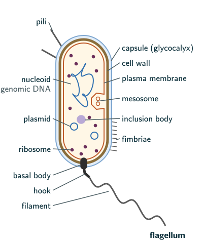
- The genomic DNA is a single chromosome, a circular DNA, lying naked in the cytoplasm (page 90). The region it occupies is called the nucleoid (beyond the NCERT text), as labelled in the figure. NCERT’s “naked” means “not enveloped by a nuclear membrane” (page 90).
- Many bacteria also have small circular DNA outside the genomic DNA, called plasmids (page 90).
- Plasmid DNA gives such bacteria certain unique phenotypic characters, one of which is resistance to antibiotics. Plasmid DNA is also used to monitor bacterial transformation with foreign DNA (page 90); NCERT returns to this in Class 12 biotechnology.
The cell envelope
Most prokaryotic cells, particularly bacterial cells, have a chemically complex cell envelope of three tightly bound layers: the outermost glycocalyx, then the cell wall, then the plasma membrane (page 90).
Although each layer of the envelope performs distinct function, they act together as a single protective unit.
NCERT Biology (Class 11), Section 8.4.1, page 90
- Glycocalyx: differs in composition and thickness among bacteria. It may be a loose sheath, the slime layer, or thick and tough, the capsule (page 90).
- Cell wall: determines the shape of the cell and gives strong structural support, preventing the bacterium from bursting or collapsing (page 90).
- Plasma membrane: selectively permeable; it interacts with the outside world and is structurally similar to that of eukaryotes (page 90).
Gram positive and Gram negative bacteria
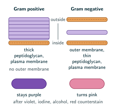
NCERT divides bacteria into two groups by differences in their cell envelopes and by how they respond to the staining procedure developed by Gram: those that take up the Gram stain are Gram positive, and those that do not are Gram negative (page 90).
Beyond the NCERT text (Wikipedia, “Gram stain”, facts in our words): the stain is named after the Danish bacteriologist Hans Christian Gram, who developed it in 1884. Today cells are stained with crystal violet, treated with iodine, which binds the dye inside the cell, washed with alcohol and then given a red counterstain such as safranin. All cells take up the violet at first; what differs is whether they keep it after the alcohol wash:
- Gram positive bacteria have a thick wall of peptidoglycan and no outer membrane; they keep the violet stain and look purple.
- Gram negative bacteria have a thin peptidoglycan layer covered by an outer membrane; the alcohol washes the violet out, so they take the counterstain and look pink.
Mesosomes and chromatophores
A specialised differentiated form of cell membrane called mesosome is the characteristic of prokaryotes.
NCERT Biology (Class 11), Section 8.4, page 90
- Mesosomes are formed by extensions of the plasma membrane into the cell, as vesicles, tubules and lamellae (page 90).
- They help in cell wall formation, DNA replication and distribution to daughter cells, respiration and secretion, and they increase the surface area of the plasma membrane and its enzymatic content (pages 90–91).
- In some prokaryotes, such as cyanobacteria, other membranous extensions into the cytoplasm, the chromatophores, contain pigments (page 91).
Beyond the NCERT text: when bacteria were later prepared for electron microscopy by rapid freezing instead of chemical fixation, mesosomes were usually not seen, and since the 1980s they have generally been regarded as artefacts of chemical fixation (Wikipedia, “Mesosome”, facts in our words). In NCERT-based exams, describe them as NCERT does.
Flagella, pili and fimbriae
- Bacterial cells may be motile or non-motile. Motile bacteria have thin filamentous extensions from their cell wall, the flagella; bacteria differ in the number and arrangement of flagella (page 91).
- A bacterial flagellum has three parts: the filament, the hook and the basal body. The filament is the longest portion and extends from the cell surface to the outside (page 91).
- Pili and fimbriae are also surface structures, but they play no role in motility. Pili are elongated tubular structures made of a special protein; fimbriae are small bristle-like fibres sprouting out of the cell. In some bacteria, fimbriae help attach the bacteria to rocks in streams and to the host tissues (page 91).
Beyond the NCERT text: a flagellum is not an outgrowth of the wall. It is a protein filament whose basal body is embedded in the plasma membrane and passes through the cell wall, as in the figure above; the basal body works as a motor that turns the filament. In NCERT-based exams, NCERT’s wording “extensions from their cell wall” is accepted.
Beyond the NCERT text: in some bacteria, type IV pili do move the cell. They attach to a surface and then retract, pulling the bacterium forward in jerky steps called twitching motility (Wikipedia, “Pilus”, facts in our words). In an NCERT-based answer, keep NCERT’s line that pili and fimbriae play no role in motility (page 91).
Ribosomes and inclusion bodies
- In prokaryotes, ribosomes are associated with the plasma membrane. They are about 15 nm by 20 nm and are made of two subunits, 50S and 30S, which together form the 70S prokaryotic ribosome (page 91).
- Several ribosomes may attach to a single mRNA and form a chain, the polyribosome or polysome; its ribosomes translate the mRNA into proteins (page 91).
Ribosomes are the site of protein synthesis.
NCERT Biology (Class 11), Section 8.4.2, page 91
Beyond the NCERT text: in bacteria such as E. coli most ribosomes are free in the cytoplasm, as in the figure; only those making membrane and secreted proteins sit on the plasma membrane. In NCERT-based exams, give NCERT’s statement.
Why is 50S + 30S not 80S? NCERT explains later that the S (Svedberg unit) is the sedimentation coefficient, indirectly a measure of density and size (Section 8.5.6, page 98). S is a settling rate, not a mass: a heavier particle settles faster, but a bigger one also meets more drag, which depends on its size and shape. So S grows more slowly than mass, and when two subunits join, their masses add but their S values do not.
- Inclusion bodies store reserve material in the cytoplasm. They are not bound by any membrane and lie free in the cytoplasm: for example, phosphate granules, cyanophycean granules and glycogen granules (page 91); glycogen is a branched polymer of glucose (NCERT Chapter 9, page 110).
- Gas vacuoles are found in blue-green, purple and green photosynthetic bacteria (page 91).
Solved examples
Example 1: Slime layer or capsule?
How does a slime layer differ from a capsule?
Answer: both are forms of the glycocalyx. A slime layer is a loose sheath; a capsule is thick and tough (NCERT page 90).
Example 2: What mesosomes do
List the functions NCERT gives for mesosomes.
Answer: cell wall formation, DNA replication and distribution to daughter cells, respiration, secretion, and increasing the surface area and enzymatic content of the plasma membrane (NCERT pages 90–91).
Example 3: Adding Svedberg units
A prokaryotic ribosome has subunits of 50S and 30S. Why is the whole ribosome 70S and not 80S?
Answer: S is a sedimentation coefficient, a settling rate that depends on density and size (NCERT page 98) and on shape; it is not a mass, so the values of the subunits do not add. The complete prokaryotic ribosome is 70S (NCERT page 91).
Example 4: Which structure moves the cell?
Of flagella, pili and fimbriae, which help a bacterium move?
Answer: only flagella; pili and fimbriae are surface structures that play no role in motility (NCERT page 91). (Beyond the NCERT text, the type IV pili of some bacteria give a twitching motility; see the note above.)
Example 5: Plasmids
Why do plasmids matter in medicine?
Answer: plasmid DNA can give bacteria resistance to antibiotics (NCERT page 90).
Common mistakes to avoid
Common mistake
Saying every prokaryote has a cell wall
Mycoplasma has no cell wall (NCERT page 90).
Common mistake
Adding the subunits to get 80S
50S + 30S form a 70S ribosome; S values are not additive. 80S is the eukaryotic ribosome, with 60S and 40S subunits (NCERT page 98).
Common mistake
Giving pili or fimbriae a role in movement
Flagella move the cell; pili and fimbriae play no role in motility (NCERT page 91). (Beyond the NCERT text, the type IV pili of some bacteria give a twitching motility; see the note in the flagella section.)
Common mistake
Thinking 70S ribosomes occur only in prokaryotes
Mitochondria (NCERT page 97) and chloroplasts (NCERT page 98) also have 70S ribosomes; the cytoplasm of eukaryotic cells has 80S ribosomes (NCERT page 98). Beyond the NCERT text, the mitochondrial ribosome of mammals is smaller, about 55S (Wikipedia, “Mitochondrial ribosome”, facts in our words); give NCERT’s 70S in an NCERT-based answer.
Common mistake
Calling inclusion bodies membrane-bound
Inclusion bodies are not bound by any membrane; they lie free in the cytoplasm (NCERT page 91).
Quick revision summary
- Prokaryotes: bacteria, blue-green algae, mycoplasma and PPLO; smaller than eukaryotic cells and multiply faster; four bacterial shapes.
- Naked circular genomic DNA, no nuclear membrane; plasmids carry traits such as antibiotic resistance.
- A cell wall in all except mycoplasma; no organelles except ribosomes; inclusions.
- Envelope: glycocalyx (slime layer or capsule), cell wall, plasma membrane, acting as one protective unit; Gram positive bacteria take up the Gram stain, Gram negative do not.
- Mesosomes: infoldings of the plasma membrane (vesicles, tubules, lamellae) for wall formation, DNA replication and distribution, respiration and secretion; chromatophores in cyanobacteria.
- Flagellum: filament, hook, basal body; pili and fimbriae are not for motility.
- 70S ribosomes (50S and 30S subunits), about 15 nm by 20 nm, in the cytoplasm and associated with the plasma membrane; polysomes; inclusion bodies (phosphate, cyanophycean, glycogen granules); gas vacuoles.
Formula sheet
- bacterial shapes: bacillus, coccus, vibrio, spirillum
- cell envelope: glycocalyx, cell wall, plasma membrane
- glycocalyx: slime layer (loose) or capsule (thick, tough)
- flagellum: filament, hook, basal body
- prokaryotic ribosome: 70S, of 50S and 30S subunits (S values do not add)
- inclusion bodies: phosphate, cyanophycean, glycogen granules
Questions students ask
Is mycoplasma Gram negative?
Mycoplasmas are not truly Gram negative, nor Gram positive, because they have no cell wall: NCERT says all prokaryotes have a cell wall surrounding the cell membrane except mycoplasma (page 90), and it sorts bacteria into the two Gram groups by differences in their cell envelopes. Beyond the NCERT text, Gram’s method sorts bacteria by whether they keep its crystal violet stain (Gram positive) or lose it (Gram negative), and MeSH (US National Library of Medicine) files the wall-less mycoplasmas as a gram-negative genus, so “Gram negative” is only a loose label for them (reasoned).
What is the difference between pili and fimbriae?
Pili are elongated tubular structures made of a special protein, while fimbriae are small bristle-like fibres sprouting out of the cell; NCERT adds that in some bacteria fimbriae help attach the bacteria to rocks in streams and to host tissues (page 91). NCERT describes both as surface structures without a motility role; beyond the NCERT text, some bacteria creep over surfaces by twitching motility, anchoring type IV pili and reeling them back in (Wikipedia, “Pilus”, facts in our words). Also beyond the NCERT text, fimbriae are short and numerous, and some longer pili, the sex pili, join two bacteria so that DNA passes from one to the other in conjugation.
What is the difference between plasmid DNA and genomic DNA?
A bacterium’s genomic DNA is its one circular chromosome, with no nuclear membrane around it, while plasmids are small circular DNA molecules outside the genomic DNA, which many bacteria, but not all, carry (NCERT page 90). Plasmid DNA confers certain unique phenotypic characters, such as resistance to antibiotics, and NCERT Class 12 calls a plasmid an autonomously replicating circular extra-chromosomal DNA, which is why plasmids are used as vectors to carry foreign DNA. Beyond the NCERT text, a bacterium can usually survive without its plasmids but not without its genomic DNA, which carries the genes for its essential functions.
Is a plasmid the same as a plastid?
A plasmid is not a plastid: a plasmid is a small circular DNA found outside the genomic DNA in many bacteria, while a plastid is a membrane-bound organelle found in all plant cells and in euglenoids (NCERT pages 90 and 97). Plastids are of three kinds, chloroplasts, chromoplasts and leucoplasts, which carry pigments or store food; the chloroplast, which traps light energy for photosynthesis, even contains its own small circular DNA. Prokaryotic cells have no plastids, since ribosomes are the only organelles they have.
Do prokaryotic cells have mitochondria and chloroplasts?
Prokaryotic cells have no mitochondria and no chloroplasts: NCERT says no organelles like those of eukaryotes are found in prokaryotic cells except ribosomes (page 90). Respiration is instead among the functions NCERT gives the mesosome, and cyanobacteria hold their pigments in chromatophores, not chloroplasts (pages 90–91). Beyond the NCERT text, most biologists now think mesosomes are artefacts, made when cells are chemically fixed for the electron microscope (Wikipedia, “Mesosome”, facts in our words), so the respiratory role NCERT gives them belongs to the plasma membrane itself (reasoned).
Practice questions
Try each question first, then open the answer to check your working.
-
Q1. Which prokaryote lacks a cell wall?
Show answerHide answer
Mycoplasma (NCERT page 90).
-
Q2. What are chromatophores, and where are they found?
Show answerHide answer
Membranous extensions into the cytoplasm that contain pigments, found in some prokaryotes such as cyanobacteria (NCERT page 91).
-
Q3. Name the longest part of a bacterial flagellum.
Show answerHide answer
The filament, which extends from the cell surface to the outside (NCERT page 91).
-
Q4. What is a polysome?
Show answerHide answer
A chain of several ribosomes attached to a single mRNA, which translate it into proteins (NCERT page 91).