Anatomy of dicot and monocot plants
NCERT Class 11 Biology Chapter 6 Section 6.2
Quick answer
In NCERT’s representative sections, a dicot root has fewer xylem patches and a small pith, while a monocot root is usually polyarch with a large pith. Typical dicot stems have a ring of open bundles, while monocot stems have scattered closed bundles. Dorsiventral leaves have palisade and spongy mesophyll; isobilateral leaves are more alike on both sides, with bulliform cells in grasses, but these class-level patterns have exceptions.
With your textbook
NCERT Biology (Class 11), Chapter 6 “Anatomy of Flowering Plants”, Section 6.2 “Anatomy of Dicotyledonous and Monocotyledonous Plants”, pages 73–77.
Open the official NCERT chapter (PDF, 1.9 MB, ncert.nic.in) (opens in a new tab)
Checked against the 2026-27 reprint on .
Studying plant anatomy
Anatomy of dicots and monocots
For a better understanding of tissue organisation of roots, stems and leaves, it is convenient to study the transverse sections of the mature zones of these organs.
NCERT Biology (Class 11), Section 6.2, page 73
- Monocotyledonous and dicotyledonous plants show marked variation in internal structure: they differ in the type, number and location of their vascular bundles (NCERT Summary, page 77).
- Secondary growth occurs in most dicotyledonous roots and stems (page 77).
In plain words: cut a root, a stem or a leaf straight across and look at the section under a microscope. The same three tissue systems (epidermal, ground and vascular) appear every time, but the arrangement of vascular bundles and leaf tissues helps identify typical dicot and monocot organs. The following are NCERT’s representative patterns, not exceptionless diagnostic rules for every species; compare several characters when deciding whether a specimen is a dicot or a monocot.
Dicotyledonous root
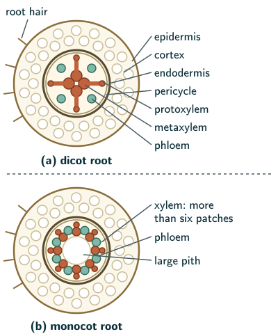
NCERT describes the transverse section of a sunflower root (pages 73–74):
- Epiblema: the outermost layer; many of its cells grow out as unicellular root hairs.
- Cortex: several layers of thin-walled parenchyma with intercellular spaces.
- Endodermis: the innermost layer of the cortex, a single layer of barrel-shaped cells without intercellular spaces. Its walls carry Casparian strips, barriers to apoplastic movement. NCERT describes them as suberin deposits; the distinction from modern chemical evidence is explained below.
- Pericycle: a few layers of thick-walled parenchyma next to the endodermis.
- Vascular bundles: usually two to four patches of xylem and of phloem, on alternating radii; the parenchyma between xylem and phloem is the conjunctive tissue. Later, a cambium ring develops between xylem and phloem.
- Xylem: radial bundles; the protoxylem lies towards the periphery (exarch), as NCERT Figure 6.3 shows; stems are endarch.
- Pith: small or inconspicuous.
- Stele: all the tissues inside the endodermis: pericycle, vascular bundles and pith.
Of the pericycle, NCERT says:
Initiation of lateral roots and vascular cambium during the secondary growth takes place in these cells.
NCERT Biology (Class 11), Section 6.2.1, page 74
NCERT says the casparian strips lie on “the tangential as well as radial walls” of the endodermal cells (page 74). Beyond the NCERT text: the casparian strip is a band running round each cell on its radial and transverse walls, the walls it shares with neighbouring endodermal cells; the tangential walls, facing the cortex and the pericycle, do not carry it. Modern qualification: Naseer and colleagues’ 2012 primary study found the Arabidopsis Casparian-strip barrier to be lignin-based, distinct from the suberin lamellae deposited later. Do not conflate the narrow strip with all endodermal suberisation; retain NCERT’s wording when specifically asked for its description. The same page also misprints parenchymatous as “parenchyomatous” and conjunctive as “conjuctive”.
Monocotyledonous root
The monocot root is similar to the dicot root in many respects: it has epidermis, cortex, endodermis, pericycle, vascular bundles and pith (page 74).
- It usually has more than six xylem bundles (polyarch), against two to four in the dicot root.
- Its pith is large and well developed.
- Monocot roots do not undergo any secondary growth (page 74).
Dicotyledonous stem
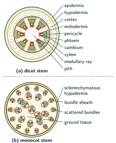
A transverse section of a young dicot stem shows (pages 74–76):
- Epidermis: the outermost protective layer, covered with a thin cuticle; it may bear trichomes and a few stomata.
- Cortex, in three sub-zones: the hypodermis, a few layers of collenchyma that give mechanical strength to the young stem; layers of rounded, thin-walled parenchyma with conspicuous intercellular spaces; and the endodermis, whose cells are rich in starch grains, so it is also called the starch sheath.
- Pericycle: on the inner side of the endodermis and above the phloem, as semi-lunar patches of sclerenchyma.
- Medullary rays: a few layers of radially placed parenchyma between the vascular bundles.
- Vascular bundles: many, arranged in a ring, the arrangement characteristic of the dicot stem.
- Pith: many rounded parenchyma cells with large intercellular spaces in the centre.
Each vascular bundle is conjoint, open, and with endarch protoxylem.
NCERT Biology (Class 11), Section 6.2.3, page 76
Monocotyledonous stem
- A sclerenchymatous hypodermis.
- Many scattered vascular bundles, each surrounded by a sclerenchymatous bundle sheath; the peripheral bundles are generally smaller than the central ones.
- A large, conspicuous parenchymatous ground tissue. Beyond the NCERT text: it is not divided into cortex, pericycle and pith, and there are no medullary rays.
- The bundles are conjoint and closed; the phloem parenchyma is absent, and water-containing cavities are present within the bundles (page 76).
Dorsiventral (dicotyledonous) leaf
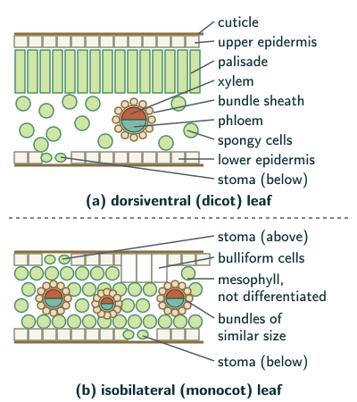
A vertical section through the lamina shows three main parts: epidermis, mesophyll and vascular system (page 76).
- Epidermis: covers both the upper (adaxial) and lower (abaxial) surfaces and has a conspicuous cuticle. The abaxial epidermis generally bears more stomata; the adaxial may even lack them.
- Mesophyll: the parenchyma between the two epidermises, with chloroplasts, which carries out photosynthesis. It has two kinds of cells: the adaxial palisade parenchyma, of elongated cells arranged vertically and parallel to each other, and below it the spongy parenchyma, of oval or round, loosely arranged cells with large spaces and air cavities.
- Vascular system: vascular bundles in the veins and the midrib, their size depending on the size of the vein; each is surrounded by a layer of thick-walled bundle sheath cells. In the reticulate venation of dicot leaves the veins vary in thickness (pages 76–77).
NCERT asks the reader to find the position of the xylem in the bundle (page 77). Beyond the NCERT text: the xylem lies towards the upper (adaxial) side and the phloem towards the lower side.
Isobilateral (monocotyledonous) leaf
The isobilateral leaf is similar to the dorsiventral leaf in many ways, with these differences (page 77):
- Stomata are present on both surfaces.
- The mesophyll is not differentiated into palisade and spongy parenchyma.
- Its parallel venation shows as vascular bundles of near similar size, except in the main veins.
Bulliform cells
In grasses, certain adaxial epidermal cells along the veins modify themselves into large, empty, colourless cells.
NCERT Biology (Class 11), Section 6.2.6, page 77
These are bulliform cells. They are living, highly vacuolated cells; NCERT’s “empty” appearance does not mean dead cells. Beyond the NCERT text: “along the veins” means in rows parallel to the veins; in section the bulliform cells sit in the upper epidermis between two veins, not over one. When they have absorbed water and are turgid, the leaf surface is exposed; when they are flaccid because of water stress, they make the leaf curl inwards, which reduces water loss (page 77).
Solved examples
Example 1: Root and stem compared (NCERT Exercise Q1)
Draw illustrations to bring out the anatomical differences between (a) a monocot root and a dicot root, and (b) a monocot stem and a dicot stem.
Answer: (a) roots:
| Feature | Dicot | Monocot |
|---|---|---|
| Xylem patches | two to four | more than six |
| Pith | small | large |
| Cambium | develops later | none |
| Secondary growth | occurs | none |
(b) stems:
| Feature | Dicot | Monocot |
|---|---|---|
| Bundles | in a ring | scattered |
| Bundle type | conjoint, open | conjoint, closed |
| Bundle sheath | none | sclerenchyma |
| Phloem parenchyma | present | absent |
| Water cavities | absent | present |
The hypodermis is collenchymatous in the dicot stem and sclerenchymatous in the monocot stem; the dicot stem has a cortex, starch sheath, pericycle, medullary rays and pith, the monocot stem one large ground tissue (NCERT pages 73–77).
Draw: a dicot root with four xylem arms and a small pith and a monocot root with eight or more and a large pith, the protoxylem outermost in both; a dicot stem with a ring of bundles, protoxylem towards the pith and medullary rays between; a monocot stem with scattered sheathed bundles, phloem facing outwards and smaller bundles near the edge. The figures on this page show each.
Example 2: Monocot or dicot stem? (NCERT Exercise Q2)
You cut a young stem across. How would you tell whether it is a monocot or a dicot stem?
Answer: look at the vascular bundles and the ground tissue. Bundles in a ring, open (with cambium), with medullary rays, under a distinct cortex with a starch sheath and around a pith, below a collenchymatous hypodermis: a dicot stem. Scattered, closed bundles, each in a sclerenchymatous sheath, in one undifferentiated ground tissue under a sclerenchymatous hypodermis: a monocot stem (NCERT pages 74–76).
Example 3: Identify the section (NCERT Exercise Q3)
A section shows (a) conjoint, scattered vascular bundles surrounded by sclerenchymatous bundle sheaths, and (b) no phloem parenchyma. What is it?
Answer: a monocot stem. Conjoint bundles rule out a root, whose bundles are radial; scattered bundles in sclerenchymatous sheaths, with no phloem parenchyma, mark a monocot stem (NCERT pages 73 and 76).
Example 4: The dorsiventral leaf (NCERT Exercise Q7)
Describe the internal structure of a dorsiventral leaf.
Answer: a vertical section shows three parts: epidermis, mesophyll and vascular system. Both epidermises have a conspicuous cuticle; the abaxial (lower) epidermis generally bears more stomata, and the adaxial may lack them. The mesophyll, which has chloroplasts and photosynthesises, is the adaxial palisade parenchyma, elongated cells arranged vertically and parallel, and below it the spongy parenchyma, loose oval or round cells with large spaces and air cavities. Vascular bundles in the veins and midrib, their size following the size of the vein, are surrounded by thick-walled bundle sheath cells (NCERT pages 76–77); the xylem faces the adaxial side and the phloem the abaxial. Label: cuticle, upper epidermis, palisade, spongy cells, bundle sheath, xylem, phloem, lower epidermis and stoma, as in the leaf figure above.
Example 5: Exarch or endarch?
Where does the protoxylem lie in a root and in a dicot stem?
Answer: in a root, towards the periphery (exarch), as NCERT Figure 6.3 shows; in a dicot stem, towards the centre (endarch protoxylem, NCERT page 76).
Common mistakes to avoid
Common mistake
Giving the monocot root two to four xylem patches
That is the dicot root; the monocot root is usually polyarch, with more than six.
Common mistake
Giving the monocot stem a cortex, pith and medullary rays
Its ground tissue is one large parenchymatous mass (NCERT page 76); beyond the NCERT text, it is not divided into cortex, pericycle and pith, and it has no medullary rays.
Common mistake
Putting palisade cells below the spongy cells
The palisade parenchyma is adaxial, under the upper epidermis; the spongy parenchyma is below it.
Quick revision summary
- Dicot root: epiblema with root hairs, parenchymatous cortex, endodermis with casparian strips, pericycle (origin of lateral roots and cambium), 2–4 xylem patches, exarch; small pith; stele inside the endodermis.
- Monocot root: same layers, more than six xylem patches (polyarch), large pith, no secondary growth.
- Dicot stem: cuticle, collenchymatous hypodermis, cortex, starch sheath, sclerenchymatous pericycle patches, a ring of conjoint, open bundles with endarch protoxylem, medullary rays, pith.
- Monocot stem: sclerenchymatous hypodermis, scattered closed bundles in sclerenchymatous sheaths, large ground tissue, no phloem parenchyma, water cavities.
- Dorsiventral leaf: more stomata below, palisade above spongy, bundles in sheaths. Isobilateral leaf: stomata on both sides, undifferentiated mesophyll, bulliform cells in grasses.
Formula sheet
- dicot root: 2–4 xylem patches, exarch, small pith
- monocot root: more than 6 (polyarch), large pith
- dicot stem: bundles in a ring, open, endarch
- monocot stem: scattered, closed, sheathed bundles
- dorsiventral leaf: palisade and spongy mesophyll
- typical isobilateral leaf: stomata both sides; bulliform cells in grasses
Questions students ask
What is the difference between a dicot root and a dicot stem?
A dicot root and a dicot stem differ most in their vascular bundles: the root has radial bundles, usually two to four patches of xylem and phloem on alternate radii, with exarch xylem, while the stem has a ring of conjoint, open bundles with endarch protoxylem and medullary rays between them. The root’s outer layer, the epiblema, bears unicellular root hairs and has no cuticle, while the stem’s epidermis has a thin cuticle and may bear trichomes and a few stomata. The stem also has a collenchymatous hypodermis below its epidermis, and the root’s endodermis carries casparian strips.
Why is a dicot leaf called dorsiventral and a monocot leaf isobilateral?
A dicot leaf is called dorsiventral because its two surfaces differ: its mesophyll is differentiated into palisade and spongy parenchyma, and its lower (abaxial) epidermis generally bears more stomata than the upper one. A monocot leaf is called isobilateral, ‘equal on both sides’, because its two surfaces are alike: stomata are present on both, and the mesophyll is not differentiated into palisade and spongy parenchyma. Beyond the NCERT text, the difference suits the way the leaves are held: most dicot leaves are spread horizontally, upper surface to the light, while many monocot leaves, such as those of grasses, stand more or less upright and are lit on both sides.
What are examples of dorsiventral and isobilateral leaves?
Dorsiventral leaves are found in most dicots, for example mango and guava, while isobilateral leaves are typical of monocots, especially grasses such as maize, wheat and sugarcane. NCERT heads its sections ‘Dorsiventral (Dicotyledonous) Leaf’ and ‘Isobilateral (Monocotyledonous) Leaf’, but the link with the two classes is general rather than exact: some dicots of dry places also have isobilateral leaves.
Why does a monocot root not show secondary growth?
A monocot root does not show secondary growth because no cambium forms in it: in a dicot root a cambium ring develops later between the xylem and the phloem, but NCERT notes that monocotyledonous roots do not undergo any secondary growth. Without the conventional vascular cambium the root cannot add secondary xylem and phloem by that mechanism. This does not mean every monocot root must remain slender: root diameter also depends on primary growth and tissue expansion.
Practice questions
Try each question first, then open the answer to check your working.
-
Q1. Why is the endodermis of a dicot stem called the starch sheath?
Show answerHide answer
Because its cells are rich in starch grains (NCERT page 75).
-
Q2. In which layer of the root do lateral roots begin?
Show answerHide answer
The pericycle (NCERT page 74).
-
Q3. What happens to a grass leaf when its bulliform cells lose water?
Show answerHide answer
The leaf curls inwards, reducing water loss (NCERT page 77).
-
Q4. Which surface of a dorsiventral leaf generally has more stomata?
Show answerHide answer
The lower (abaxial) surface (NCERT page 76).