Neuron, nerve impulse and synapse
NCERT Class 11 Biology Chapter 18 Section 18.3
Quick answer
A neuron receives and transmits signals; a typical neuron has a cell body, dendrites and an axon. Threshold depolarisation triggers a regenerative action potential through voltage-gated ion channels. At a chemical synapse, presynaptic calcium entry triggers neurotransmitter release, and the next cell’s response depends on its receptors and combined inputs.
With your textbook
NCERT Biology (Class 11), Chapter 18 “Neural Control and Coordination”, Section 18.3 “Neuron as Structural and Functional Unit of Neural System”, pages 231–235.
Open the official NCERT chapter (PDF, 4.3 MB, ncert.nic.in) (opens in a new tab)
Checked against the 2026-27 reprint on .
Neuron
Neuron
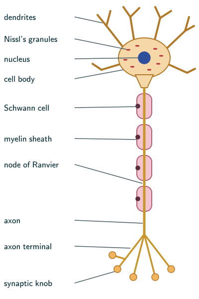
NCERT calls the neuron the structural and functional unit of the neural system, made of three major parts: the cell body, the dendrites and the axon (Section 18.3, page 231). Why it can carry signals at all:
Neurons are excitable cells because their membranes are in a polarised state.
NCERT Biology (Class 11), Section 18.3.1, page 232
In plain words: a neuron is a cell built to pass on messages. The dendrites are its receiving branches, the cell body is the hub, and the axon is a long cable that carries the message away. An action potential is a brief, regenerative change in voltage across the axon membrane. At a chemical synapse, neurotransmitter passes the signal to the next cell; electrical synapses instead pass current through connecting channels. This is the standard neuron model, not the shape of every neuron.
Cell body, dendrites and axon
NCERT describes the three parts (pages 231–232; see neural tissue):
- Cell body: cytoplasm with typical cell organelles and granular bodies called Nissl’s granules.
- Dendrites: short fibres that branch repeatedly and project out of the cell body. They also contain Nissl’s granules, and they carry impulses towards the cell body.
- Axon: a long fibre whose distal end is branched. Each branch ends in a bulb-like synaptic knob, which holds synaptic vesicles filled with chemicals called neurotransmitters. Axons carry impulses away from the cell body, to a synapse or to a neuromuscular junction.
Multipolar, bipolar and unipolar neurons
NCERT sorts neurons by the number of axons and dendrites (page 232):
- Multipolar: one axon and two or more dendrites; found in the cerebral cortex.
- Bipolar: one axon and one dendrite; found in the retina of the eye.
- Unipolar: a cell body with one axon only; found usually in the embryonic stage.
Myelinated and non-myelinated fibres
- In the PNS, myelinated fibres are enveloped by Schwann cells, which form a myelin sheath around the axon. The gaps between two adjacent myelin sheaths are the nodes of Ranvier. Myelinated fibres are found in spinal and cranial nerves (NCERT page 232).
- A peripheral unmyelinated fibre can be enclosed by a Schwann cell that does not form a myelin sheath around the axon. Such fibres are common in the autonomic and the somatic neural systems (page 232). NCERT prints “autonomous” here; it means the autonomic neural system of Section 18.2.
- Beyond the NCERT text, Schwann cells make the myelin outside the brain and spinal cord; inside them, “oligodendrocytes produce myelin”. The same module notes that “Myelinated fibers make up the white matter in the CNS, while cell bodies and unmyelinated fibers make the gray matter” (NCI SEER Training Modules, Nerve Tissue).
- Myelin also speeds the impulse. Beyond the NCERT text, a 2020 study puts it this way: “The propagation of electrical impulses along axons is highly accelerated by the myelin sheath and produces saltating or ‘jumping’ action potentials across internodes, from one node of Ranvier to the next” (Cohen and colleagues, Cell, 2020, CC BY 4.0).
Resting potential
Why is a resting neuron’s membrane polarised? NCERT builds the answer in steps (pages 232–233):
- The neural membrane has different ion channels, each selectively permeable to different ions.
- At rest, the axonal membrane is comparatively more permeable to K and nearly impermeable to Na. It is impermeable to the negatively charged proteins in the axoplasm.
- The axoplasm holds a high concentration of K and negatively charged proteins and a low concentration of Na, while the fluid outside has low K and high Na: a concentration gradient.
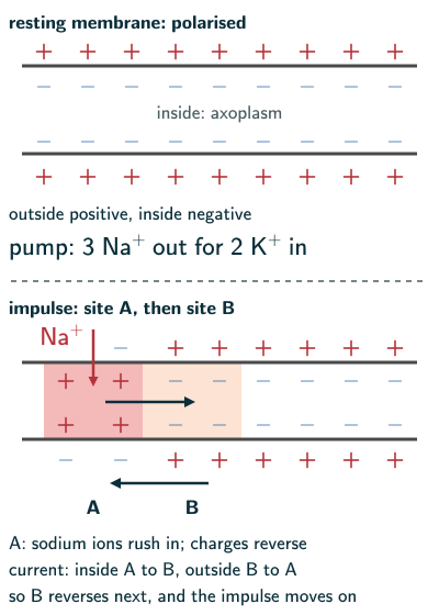
- The sodium-potassium pump keeps these gradients by active transport, moving 3 Na outwards for 2 K into the cell.
- K tending to leave through resting channels, together with unequal ion concentrations and selective permeability, makes the inside negative relative to the outside. The pump maintains the gradients over time and also contributes a smaller direct electrogenic effect. The voltage difference is the resting potential (NCERT pages 232–233; Purves and colleagues, Neuroscience, “How Ionic Movements Produce Electrical Signals”). This is a tiny charge separation near the membrane, not a bulk solution full of uncompensated charge.
Action potential and its conduction
NCERT Section 18.3.1 (page 233), with sites A and B of its Figure 18.2, supplemented with the threshold/channel mechanism:
- Sufficient depolarisation at site A reaches threshold, opening voltage-gated Na channels and sharply increasing Na permeability. A weak, subthreshold stimulus does not automatically trigger an action potential.
- A rapid influx of Na reverses the polarity at A: the outer surface becomes negative and the inner side positive. The membrane at A is depolarised.
- This rapid, regenerative voltage change is the action potential, or nerve impulse. Depolarisation means becoming less negative; reversal to a positive value is the overshoot of this action potential, not the definition of every depolarisation.
- Just ahead, at site B, the membrane is still positive outside and negative inside. So a current flows on the inner surface from A to B, and on the outer surface from B to A, completing the circuit.
- Local current depolarises B to threshold, opening its voltage-gated channels and regenerating the action potential there. The sequence repeats along the axon, and the impulse is conducted.
- Na channels inactivate, while increased K permeability allows K to leave, producing repolarisation. The membrane may briefly become more negative than at rest. Recovery of channel availability creates absolute and relative refractory periods; it is not simply the pump immediately restoring all ion concentrations after each impulse (Purves and colleagues, Neuroscience; StatPearls, “Physiology, Action Potential”).
NCERT’s Summary puts the whole run in one line: the nerve impulse is conducted along the axon membrane “in the form of a wave of depolarisation and repolarisation” (page 237).
Electrical and chemical synapses
A nerve impulse passes from one neuron to the next through junctions called synapses (NCERT Section 18.3.2, page 234). A synapse is formed by the membranes of a pre-synaptic and a post-synaptic neuron, with the prominent synaptic cleft characteristic of chemical synapses. Electrical synapses also have a very narrow intercellular gap, bridged by gap-junction channels; the cell membranes do not fuse (Purves and colleagues, Neuroscience, “Electrical Synapses”). There are two types (page 234):
- Electrical synapses: the two membranes are in very close proximity, and current flows directly through gap-junction channels from one neuron into the other. Transmission is very similar to conduction along a single axon with very little synaptic delay; NCERT describes it as always faster than chemical transmission. NCERT calls electrical synapses rare in our system, meaning much less prominent in its account than chemical synapses; they nevertheless occur in important mammalian circuits.
- Chemical synapses: the two membranes are separated by a fluid-filled synaptic cleft, and chemicals called neurotransmitters carry the impulse across.
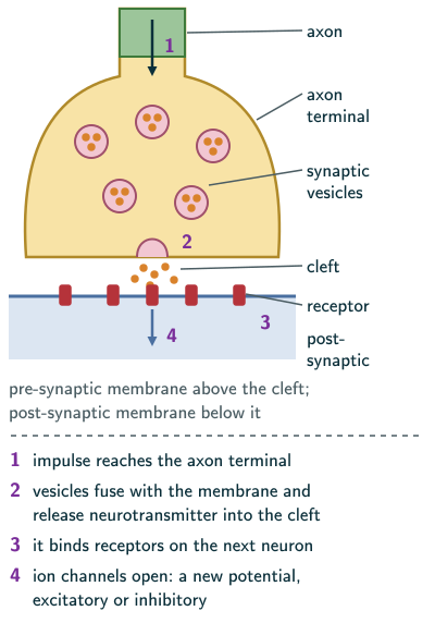
At a chemical synapse, transmission runs in four steps (pages 234–235):
- The axon terminals contain vesicles filled with neurotransmitters.
- The arriving impulse opens voltage-gated Ca channels; Ca entry triggers fusion of release-ready synaptic vesicles and neurotransmitter release into the cleft. NCERT describes vesicle movement/fusion; calcium supplies the key mechanistic link (Purves and colleagues, Neuroscience, “Second Messengers”).
- The neurotransmitters bind to specific receptors on the post-synaptic membrane.
- Receptors change postsynaptic ion-channel activity, directly or through signalling pathways. The resulting graded postsynaptic potential may be excitatory or inhibitory; ion movement can be inward or outward, depending on the channel and electrochemical gradient. An excitatory potential increases the chance of firing, but does not guarantee a new action potential: the combined inputs must reach threshold (Purves and colleagues, Neuroscience, “Excitatory and Inhibitory Postsynaptic Potentials”).
One neurotransmitter met elsewhere in the book is acetylcholine, released at the neuromuscular junction to start muscle contraction (NCERT Chapter 17, page 222).
Solved examples
Example 1: Resting and action potential (NCERT Exercise 2b)
Compare the resting potential and the action potential.
Answer: the resting potential is the electrical potential difference across the membrane of a resting neuron, when the outer surface is positive and the inner surface negative (polarised), with high K inside and high Na outside kept by the sodium-potassium pump. The action potential is a brief regenerative voltage change after threshold is reached: a rapid influx of Na has reversed the polarity, so that the outside is negative and the inside positive (depolarised); it is the nerve impulse, and it lasts only a fraction of a second before K diffuses out and restores the resting potential (NCERT pages 232–233).
Example 2: Polarisation and depolarisation (NCERT Exercise 3a, b)
Explain (a) polarisation and (b) depolarisation of the membrane of a nerve fibre.
Answer: (a) At rest the membrane is more permeable to K, nearly impermeable to Na and impermeable to the negatively charged proteins of the axoplasm, and the sodium-potassium pump sends 3 Na out for every 2 K in. So the outer surface is positive and the inner surface negative: the membrane is polarised. (b) A stimulus that brings the membrane to threshold opens voltage-gated Na channels; Na rushes in and the polarity reverses, outside negative and inside positive: the membrane is depolarised (NCERT pages 232–233).
Example 3: The role of Na (NCERT Exercise 7)
Explain the role of Na in the generation of an action potential.
Answer: at rest Na is kept high outside the axon, because the membrane is nearly impermeable to it and the pump moves it out. Sufficient depolarisation reaches threshold and opens voltage-gated Na channels, and the rapid influx of Na reverses the polarity at that site: this reversal is the action potential (NCERT pages 232–233).
Example 4: Synaptic transmission (NCERT Exercises 3c, 5e and 6)
Give a brief account of how an impulse crosses a chemical synapse.
Answer: at a chemical synapse, an arriving action potential opens presynaptic voltage-gated Ca channels. Calcium entry triggers vesicle fusion and neurotransmitter release into the fluid-filled cleft. The transmitter binds postsynaptic receptors and changes ion-channel activity, producing an excitatory or inhibitory graded response. The postsynaptic neuron fires an action potential only if its integrated inputs reach threshold (NCERT pages 234–235; the calcium and threshold detail is explained above).
Example 5: Myelinated and non-myelinated axons (NCERT Exercises 8a and 10b)
Distinguish between myelinated and non-myelinated axons, including how they conduct an impulse.
Answer: in the PNS, a myelinated axon is wrapped by Schwann cells that form a myelin sheath, broken by nodes of Ranvier; such fibres are found in spinal and cranial nerves. A non-myelinated axon is enclosed by a Schwann cell that forms no myelin sheath; such fibres are common in the autonomic and somatic neural systems (NCERT page 232). In a non-myelinated fibre, the impulse is conducted as NCERT describes on page 233: the action potential at one site reverses the polarity of the site just ahead, and the sequence is repeated along the whole length of the axon. Beyond the NCERT text, myelin makes conduction much faster: local current spreads rapidly beneath myelin and action potentials are regenerated at successive nodes of Ranvier, called saltatory conduction (Cohen and colleagues, Cell, 2020).
Example 6: Dendrites and axons (NCERT Exercise 8b)
Differentiate between dendrites and axons.
Answer: dendrites are short fibres that branch repeatedly, contain Nissl’s granules and carry impulses towards the cell body. The axon is a long fibre, branched at its distal end into synaptic knobs with neurotransmitter vesicles, and it carries impulses away from the cell body to a synapse or a neuromuscular junction (NCERT pages 231–232).
Example 7: Counting the pump
Using NCERT’s ratio of 3 Na out for 2 K in, how many Na and K ions move in 50 cycles of the pump, and how many more positive charges leave the cell than enter it?
Answer: 50 × 3 = 150 Na out and 50 × 2 = 100 K in, so 150 − 100 = 50 more positive charges leave than enter, one per cycle (from NCERT page 232). NCERT’s point is that the pump maintains the ionic gradients across the resting membrane (page 232).
Example 8: Drawing a neuron (NCERT Exercise 4a)
Which labels should a diagram of a neuron carry?
Answer: dendrites, cell body, nucleus, Nissl’s granules, axon, Schwann cell, myelin sheath, node of Ranvier, axon terminal and synaptic knob, as in the figure above (NCERT Figure 18.1, page 232).
Common mistakes to avoid
Common mistake
Sending impulses the wrong way along dendrites
Dendrites carry impulses towards the cell body; the axon carries them away from it (NCERT page 232).
Common mistake
Making the inside of a resting axon positive
At rest the outer surface is positive and the inner surface negative; an action potential briefly reverses that local voltage difference (NCERT pages 232–233).
Common mistake
Letting Na flow out during depolarisation
In depolarisation Na rushes in; it is K that then diffuses out to restore the resting potential (NCERT page 233).
Common mistake
Calling electrical synapses slower
NCERT describes electrical transmission as faster because current passes directly between cells with little delay (page 234). This does not mean electrical synapses lack a junctional gap: gap-junction channels bridge it.
Quick revision summary
- A neuron has a cell body with Nissl’s granules, dendrites that carry impulses towards it, and an axon that carries them away to synaptic knobs holding neurotransmitter vesicles.
- Multipolar neurons (cerebral cortex), bipolar (retina), unipolar (usually the embryonic stage).
- Peripheral myelinated fibres have Schwann cells forming a myelin sheath with nodes of Ranvier (spinal and cranial nerves); unmyelinated fibres lack the sheath.
- Resting membrane: positive outside, negative inside; more permeable to K; the sodium-potassium pump moves 3 Na out for 2 K in.
- A threshold-reaching stimulus opens Na channels and Na influx reverses the local polarity: the action potential, conducted as a wave of depolarisation and repolarisation; K leaving restores the resting potential.
- Electrical synapses pass current directly and faster but are rare; chemical synapses use neurotransmitters, receptors and ion channels.
Formula sheet
- neuron: dendrites (towards the cell body) → cell body → axon (away)
- resting: outside positive, inside negative; K high inside, Na high outside
- sodium-potassium pump: 3 Na out for 2 K in
- action potential: Na in → polarity reversed; K out → resting potential back
- chemical synapse: impulse → Ca entry → vesicle fusion → transmitter → postsynaptic response
Questions students ask
Why is the resting potential of a neuron negative?
The resting potential of a neuron is negative because, at rest, the inside of the axon is negatively charged relative to the outside: NCERT explains this polarised state by a membrane comparatively more permeable to K, nearly impermeable to Na and impermeable to the axoplasm’s negatively charged proteins, with a sodium-potassium pump that transports 3 Na outwards for 2 K into the cell (NCERT pages 232–233). Beyond the NCERT text, a membrane potential is the voltage inside minus the voltage outside, so the resting value is negative: between −30 and −100 mV in excitable cells (MeSH, “Membrane Potentials”) and often illustrated as about −70 mV for a mammalian neuron, though values vary by cell type and state (Johnson and colleagues, PLOS ONE, 2026).
Why is conventional chemical synaptic transmission one-way?
A nerve impulse crosses a chemical synapse in only one direction because the two sides of the synapse are different: the vesicles filled with neurotransmitters are in the axon terminal of the pre-synaptic neuron, while the specific receptors that bind the neurotransmitters are on the post-synaptic membrane (NCERT pages 234–235). This explains the direction of conventional fast synaptic transmission. It is not a claim that neurons cannot also exchange slower feedback or retrograde signals.
Why does an action potential normally propagate forwards along an axon?
During normal forward propagation, recently active membrane behind the impulse is refractory, largely because voltage-gated Na channels are inactivated; membrane ahead is still excitable. This favours forward propagation (NCERT page 233; StatPearls, “Physiology, Action Potential”). The absolute refractory period prevents another action potential; during the relative period, a stronger stimulus can succeed. Axons are not physically one-way wires: experimentally stimulating the middle of a resting axon can launch impulses in both directions (Grosberg and colleagues, Journal of Neurophysiology, 2017).
What is the difference between a neuron and a nerve?
A neuron is a single cell, made of a cell body, dendrites and an axon (NCERT page 231), while a nerve, beyond the NCERT text, is a bundle of axons with supporting cells and connective-tissue coverings (NCI SEER Training Modules, Peripheral Nervous System; MeSH, “Peripheral Nerves”). NCERT’s PNS is all the nerves of the body associated with the CNS (page 231), and nerves lie outside the brain and spinal cord (MeSH). Inside the brain, a bundle of fibres is what NCERT calls a tract, such as the corpus callosum (page 236).
What happens when the myelin sheath is damaged?
Beyond the NCERT text, when the myelin sheath is damaged, the axon loses the insulation that speeds its impulses and communication between neurons suffers: myelin is an electrical insulator that makes conduction faster and more energy-efficient (MeSH, “Myelin Sheath”). A well-known example is multiple sclerosis, in which the immune system attacks the myelin of the brain and spinal cord, leaving scar-like patches, with symptoms such as vision loss, weakness and numbness (NINDS, Multiple Sclerosis; MeSH, “Multiple Sclerosis”). That myelin is made by oligodendrocytes (MeSH, “Oligodendroglia”), not by the Schwann cells that NCERT describes in spinal and cranial nerves (page 232).
Practice questions
Try each question first, then open the answer to check your working.
-
Q1. Where are bipolar neurons found, according to NCERT?
Show answerHide answer
In the retina of the eye (NCERT page 232).
-
Q2. What are the gaps between two adjacent myelin sheaths called?
Show answerHide answer
Nodes of Ranvier (NCERT page 232).
-
Q3. What restores the resting potential after an action potential?
Show answerHide answer
Na-channel inactivation and increased K permeability lead to repolarisation as K leaves (NCERT page 233 describes the K efflux). Refractory recovery also requires sodium channels to become available again; the pump maintains ion gradients over time.