Muscle: structure and mechanism of contraction
NCERT Class 11 Biology Chapter 17 Section 17.2
Quick answer
Skeletal muscle produces force as ATP-powered myosin heads interact with actin; calcium binding to troponin enables this interaction. During shortening, sarcomeres shorten, I bands and H zones narrow, and A bands retain their length. ATP also detaches myosin and powers calcium uptake during relaxation.
With your textbook
NCERT Biology (Class 11), Chapter 17 “Locomotion and Movement”, Section 17.2 “Muscle”, pages 218–223.
Open the official NCERT chapter (PDF, 4.5 MB, ncert.nic.in) (opens in a new tab)
Checked against the 2026-27 reprint on .
Muscle
Muscle
Muscles make up a large part of the body and do its moving. NCERT begins:
Muscle is a specialised tissue of mesodermal origin.
NCERT Biology (Class 11), Section 17.2, page 218
In plain words: a skeletal muscle is a bundle of bundles. Its long cells contain myofibrils made of repeating sarcomeres, where myosin pulls on actin to produce force. When the muscle shortens, these units shorten together. Smooth muscle shows no striations (NCERT page 218); the detailed mechanism below is for skeletal muscle.
Skeletal, visceral and cardiac muscles
- Muscles contribute about 40–50 per cent of the body weight of an adult human. They have the special properties of excitability, contractility, extensibility and elasticity (NCERT page 218).
- They are classified by location, appearance and nature of regulation. By location there are three types (page 218; see muscle tissue):
- Skeletal muscles: closely associated with the skeletal components; striped under the microscope, so striated; under voluntary control of the nervous system, so voluntary. They are mainly for locomotion and changes of body posture (page 218).
- Visceral muscles: in the inner walls of hollow visceral organs such as the alimentary canal and reproductive tract; no striations, so smooth (non-striated); not under voluntary control, so involuntary. They move food through the digestive tract and gametes through the genital tract (page 218).
- Cardiac muscles: the muscles of the heart; many cells assemble in a branching pattern; striated but involuntary. Cardiac pacemaker cells initiate the rhythm; autonomic nerves modify rate and force rather than voluntarily triggering each beat (NCERT page 219; Chapter 15, regulation of cardiac activity).
Structure of a skeletal muscle fibre
- Each skeletal muscle is made of muscle bundles (fascicles) held together by a collagenous connective tissue layer, the fascia. Each bundle contains many muscle fibres (NCERT page 219).
- Each muscle fibre is lined by the plasma membrane, the sarcolemma, enclosing the sarcoplasm. A muscle fibre is a syncytium, because its sarcoplasm contains many nuclei (page 219; NCERT prints “syncitium”).
- The endoplasmic reticulum of the fibre, the sarcoplasmic reticulum, is the store house of calcium ions (page 219).
- The sarcoplasm has a large number of parallel threads, the myofibrils, each with alternate dark and light bands (page 219).
- NCERT’s Summary calls the muscle fibre the anatomical unit of muscle; the sarcomere is the functional unit (page 228).
- NCERT calls these threads “myofilaments or myofibrils” (page 219), but the two are not the same thing. Beyond the NCERT text, each myofibril is made of many thin and thick myofilaments, of actin and myosin, which repeat as sarcomeres along its length (Zhao and colleagues, “Architecture and molecular machinery of skeletal myofibers”, Frontiers in Cell and Developmental Biology, 2025). NCERT’s own Summary keeps them apart: each muscle fibre has many myofibrils, and the thin and thick filaments make up the bands of each sarcomere (page 228).
Sarcomere
- The striations come from the arrangement of two proteins, actin and myosin (NCERT page 219):
- the light band, the I band (isotropic), contains actin;
- the dark band, the A band (anisotropic), contains myosin.
- Actin filaments are thinner than myosin filaments, so they are called thin and thick filaments. Both run parallel to each other and to the long axis of the myofibril (page 220).
- A Z line bisects each I band, and the thin filaments are firmly attached to it. An M line holds the thick filaments together in the middle of the A band (page 220).
- The part of a myofibril between two successive Z lines is the sarcomere, the functional unit of contraction (page 220).
- At rest, the edges of the thin filaments partly overlap the free ends of the thick filaments. The central part of the thick filaments, not overlapped, is the H zone (page 220).
- So the A band, the whole length of the thick filaments, also holds thin filaments near each end, where the two overlap. Only the H zone has thick filaments alone, and only the I band has thin filaments alone (NCERT page 220, Figure 17.2). Beyond the NCERT text, a 2025 review puts it the same way: “the myosins are assembled into thick myofilaments in the A-band that overlap with thin myofilaments” (Zhao and colleagues, Frontiers in Cell and Developmental Biology, 2025, CC BY 4.0).
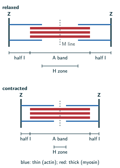
NCERT Exercise 1 asks you to draw a sarcomere and show its regions. Draw the relaxed panel above and label a Z line at each end, a half I band (thin filaments only) beside each Z line, the A band (the whole length of the thick filaments) in the middle, the H zone at the centre of the A band, and the M line through the H zone (NCERT page 220, Figure 17.2).
Structure of contractile proteins
NCERT Section 17.2.1 (page 221):
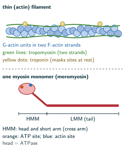
- Actin (thin) filament: two F (filamentous) actins wound helically round each other; each F actin is a polymer of G (globular) actins.
- Two filaments of tropomyosin run close to the F actins along their length.
- A complex protein, troponin, sits at regular intervals on the tropomyosin; at rest, NCERT says a subunit of troponin masks the active binding sites for myosin on actin. Beyond the NCERT text (Wikipedia, “Muscle contraction”, facts in our words): tropomyosin itself covers part of each binding site, and when Ca binds troponin C, the troponin–calcium complex moves the tropomyosin aside.
- Myosin (thick) filament: an assembly of myosin molecules. NCERT calls the monomers meromyosins and describes each using these regions:
- a globular head with a short arm, the heavy meromyosin (HMM);
- a tail, the light meromyosin (LMM).
- The HMM parts project outwards at regular distances and angles from the thick filament as cross arms. The globular head is an ATPase enzyme, with binding sites for ATP and active sites for actin.
Beyond the NCERT text: a myosin II molecule has two heads and a long tail in which the two heavy chains coil round each other, and in muscle the tails of many such molecules join to form the thick filament (Wikipedia, “Myosin”, facts in our words). HMM and LMM are the two parts obtained when myosin is cut with the enzyme trypsin (Wikipedia, “Meromyosin”, facts in our words), so they are not separate units from which a thick filament is built (our reasoning). Use NCERT’s labelled regions for its diagram questions.
Mechanism of muscle contraction
NCERT Section 17.2.2 (pages 222–223):
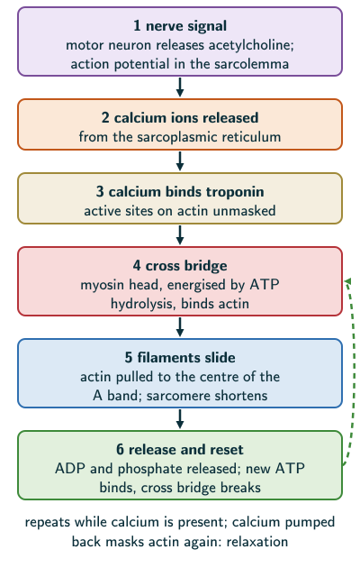
- A signal from the central nervous system arrives by a motor neuron. A motor neuron with the muscle fibres it connects to is a motor unit; its junction with the sarcolemma is the neuromuscular junction, or motor-end plate.
- The signal releases a neurotransmitter, acetylcholine, which generates an action potential in the sarcolemma. This spreads through the fibre and releases calcium ions into the sarcoplasm.
- Ca binds a subunit of troponin, removing the mask from the active sites for myosin on actin.
- Using energy from ATP hydrolysis, the myosin head binds the exposed sites on actin, forming a cross bridge.
- This pulls the actin filaments towards the centre of the A band. The Z lines attached to them are pulled inwards too, so the sarcomere shortens: contraction. During contraction, the I bands shorten while the A bands keep their length.
- Releasing ADP and P, the myosin returns to its relaxed state; a new ATP binds and the cross bridge breaks.
- The ATP is hydrolysed again and the cycle repeats, causing further sliding, until the Ca is pumped back to the sarcoplasmic cisternae. Actin is masked again, the Z lines return, and the muscle relaxes.
NCERT slip (page 222): NCERT prints “ADP and P”, with a subscript 1; it means P, inorganic phosphate, the partner of ADP when ATP is split (compare NCERT Chapter 12, page 160: ATP is made “from ADP and inorganic phosphate”). Write ADP and P in an answer.
Beyond the NCERT text (Wikipedia, “Muscle contraction”, facts in our words): in the modern account of the cycle, the head sheds its phosphate as the power stroke begins and its ADP when the stroke is over, then stays locked to actin (the rigor state) until a fresh ATP frees it; relaxation follows as a calcium pump of the sarcoplasmic reticulum (SERCA) takes Ca back. Force can also be produced with no change in length, an isometric contraction, for example when a heavy bag is held steady; NCERT’s sliding diagrams show the shortening case.
Sliding filament theory
NCERT states the theory (page 221):
Mechanism of muscle contraction is best explained by the sliding filament theory which states that contraction of a muscle fibre takes place by the sliding of the thin filaments over the thick filaments.
NCERT Biology (Class 11), Section 17.2.2, page 221
- During shortening the thin filaments slide inwards, so the I bands and the H zone become shorter, while the A band keeps its length, and the thin and thick filaments keep theirs (NCERT pages 222–223, Figure 17.5).
Red and white muscle fibres
- The reaction time of fibres varies between muscles. NCERT links repeated activation, anaerobic glycogen breakdown and lactic-acid accumulation with fatigue (page 223; see lactic acid fermentation). Beyond the NCERT text: muscle fatigue was once blamed on a build-up of lactic acid, and whether lactic acid worsens fatigue or eases it is not settled; a build-up of other metabolites can bring on fatigue within the muscle fibres by cutting the release of calcium from the sarcoplasmic reticulum or by making actin and myosin less sensitive to calcium (Wikipedia, “Muscle fatigue”, facts in our words). In an NCERT-based answer, give NCERT’s lactic-acid account.
- Muscle contains a red, oxygen-storing pigment, myoglobin (page 223).
- Red fibres: high myoglobin, so reddish; plenty of mitochondria that use the stored oxygen for ATP; they can be called aerobic muscles (page 223).
- White fibres: very little myoglobin, so pale; few mitochondria, but a lot of sarcoplasmic reticulum; they depend on anaerobic processes for energy (page 223). Red and white differ in which pathway they lean on more; neither fibre type uses only one energy pathway, since white fibres still have a few mitochondria and NCERT puts anaerobic glycogen breakdown in muscles generally (reasoned from NCERT page 223).
Solved examples
Example 1: Sliding filament theory (NCERT Exercise 2)
Define the sliding filament theory of muscle contraction.
Answer: it states that the contraction of a muscle fibre takes place by the sliding of the thin (actin) filaments over the thick (myosin) filaments (NCERT page 221).
Example 2: Steps in contraction (NCERT Exercise 3)
Describe the important steps in muscle contraction.
Answer: a signal from the CNS reaches the neuromuscular junction by a motor neuron and releases acetylcholine, which generates an action potential in the sarcolemma. Calcium ions are released into the sarcoplasm and bind a troponin subunit, unmasking the active sites on actin. Using energy from ATP hydrolysis, myosin heads bind actin as cross bridges and pull the thin filaments towards the centre of the A band, so the Z lines move in and the sarcomere shortens. The myosin releases ADP and P, a new ATP binds and the cross bridge breaks; ATP is hydrolysed again and the cycle repeats. When the Ca is pumped back into the sarcoplasmic cisternae, actin is masked again and the muscle relaxes (NCERT pages 222–223). Beyond the NCERT text, the modern order of the cycle is given above (Wikipedia, “Muscle contraction”, facts in our words).
Example 3: True or false? (NCERT Exercise 4a, b)
(a) Actin is present in the thin filament. (b) The H zone of a striated muscle fibre represents both thick and thin filaments.
Answer: (a) true (NCERT page 220). (b) False: the H zone is the central part of the thick filaments that is not overlapped by thin filaments, so it has thick filaments only (page 220).
Example 4: Differences (NCERT Exercise 5a, b)
Write the differences between (a) actin and myosin, (b) red and white muscles.
Answer: (a) Actin forms the thin filaments: two F actins, each a polymer of G actins, with tropomyosin and troponin. They are attached to the Z lines and make up the light I band, and their ends reach into the A band. Myosin forms the thick filaments of the dark A band, held together at the M line; each is an assembly of myosin molecules (NCERT calls its monomers meromyosins), whose heads are ATPases with sites for ATP and actin (NCERT pages 219–221). (b) Red fibres have much myoglobin and many mitochondria and work aerobically; white fibres have little myoglobin, few mitochondria and much sarcoplasmic reticulum, and depend on anaerobic processes (page 223).
Example 5: Match the columns (NCERT Exercise 6a–c)
Match: (a) smooth muscle, (b) tropomyosin, (c) red muscle with (i) myoglobin, (ii) thin filament, (iv) involuntary.
Answer: (a)–(iv), (b)–(ii), (c)–(i) (NCERT pages 218, 221 and 223). The fourth pair, skull–sutures, is on the joints page.
Example 6: Skeletal against cardiac muscle (NCERT Exercise 8)
How do you distinguish between a skeletal muscle and a cardiac muscle?
Answer: both are striated, but skeletal muscle is attached to skeletal components and is voluntary, while cardiac muscle is found only in the heart, has cells assembled in a branching pattern, and is involuntary. Its pacemaker initiates the rhythm, while autonomic nerves modify cardiac activity (NCERT pages 218–219; Chapter 15).
Example 7: Fill in the blanks (NCERT Exercise 10c, d)
(c) The thin filament contains 2 F actins and two other proteins, ___ and ___. (d) In a muscle fibre, Ca is stored in ___.
Answer: (c) tropomyosin and troponin (NCERT page 221); (d) the sarcoplasmic reticulum (page 219).
Common mistakes to avoid
Common mistake
Saying the A band shortens in contraction
The A band keeps its length; the I bands and the H zone shorten as the thin filaments slide in. The filaments themselves do not shorten: they slide (NCERT pages 221–223). At maximal contraction the thin filaments from both sides meet and overlap in the middle, so the H zone is lost (NCERT Figure 17.5, last panel, page 223).
Common mistake
Saying the A band has myosin only
The A band is the whole length of the thick filaments, so near its ends the thin actin filaments overlap them. Only the H zone has thick filaments alone; the I band has thin filaments alone (NCERT pages 219–220).
Common mistake
Putting both filaments in the H zone
The H zone is the part of the thick filaments not overlapped by thin filaments (NCERT page 220).
Common mistake
Letting calcium bind myosin
Ca binds a subunit of troponin on the thin filament, which unmasks the active sites on actin (NCERT page 222).
Common mistake
Thinking ATP is needed only to make the cross bridge
ATP is hydrolysed to energise the myosin head before it binds actin, and a new ATP must bind before the cross bridge can break (NCERT page 222). So ATP is needed both to pull and to let go. Beyond the NCERT text: the pump that takes Ca back into the sarcoplasmic reticulum, SERCA, is a calcium-ATPase, so relaxation uses ATP too (Wikipedia, “Muscle contraction”, facts in our words).
Common mistake
Giving red fibres more sarcoplasmic reticulum
Red fibres have more myoglobin and more mitochondria and work aerobically; white fibres have little myoglobin and few mitochondria but much more sarcoplasmic reticulum, and depend on anaerobic processes (NCERT page 223).
Quick revision summary
- Muscle is mesodermal; it is 40–50 per cent of adult body weight. Skeletal muscle is striated and voluntary, visceral muscle smooth and involuntary, cardiac muscle striated, branched and involuntary.
- Muscle → fascicles (in fascia) → muscle fibres (sarcolemma, many nuclei, sarcoplasmic reticulum storing Ca) → myofibrils of alternating I and A bands.
- Sarcomere (Z line to Z line) is the functional unit: thin actin filaments from the Z lines, thick myosin filaments of the A band held at the M line; the H zone is the unoverlapped middle of the thick filaments.
- Thin filament: two F actins (of G actin), tropomyosin, troponin. Thick filament: myosin molecules with ATPase heads; NCERT describes head/short arm (HMM) and tail (LMM). Beyond the NCERT text: HMM and LMM are the parts obtained when myosin is cut with trypsin (Wikipedia, “Meromyosin”).
- Shortening contraction: nerve signal → acetylcholine → Ca → troponin → cross bridges → thin filaments slide in; I bands and H zone shorten, A band unchanged; Ca pumped back → relaxation.
- Red fibres: myoglobin, many mitochondria, aerobic. White fibres: little myoglobin, much sarcoplasmic reticulum, anaerobic.
Formula sheet
- sarcomere: Z line → Z line (functional unit)
- shortening: I band and H zone shorten; A band unchanged
- thin filament: 2 F-actin + tropomyosin + troponin
- NCERT myosin regions: head + short arm (HMM), tail (LMM)
- trigger: acetylcholine → Ca → troponin → cross bridge
Questions students ask
Why is calcium needed for muscle contraction?
Calcium is needed for muscle contraction because it is the switch that lets myosin reach actin: in a resting fibre a subunit of troponin masks the active binding sites for myosin on the actin filaments, and Ca binding to that subunit removes the mask, so that myosin heads can form cross bridges (NCERT pages 221–222). The Ca comes from the fibre’s own store, the sarcoplasmic reticulum, released into the sarcoplasm as the action potential spreads (pages 219 and 222). When it is pumped back into the sarcoplasmic cisternae, actin is masked again and the muscle relaxes (page 223). Beyond the NCERT text, the calcium-binding part of the troponin complex is troponin C (MeSH, “Troponin C”).
Why do the I band and H zone shorten during contraction but the A band does not?
The A band keeps its length because it spans the whole length of the thick myosin filaments, which do not change, while the I band and the H zone are the parts where thin and thick filaments do not overlap, and these shrink as the thin filaments slide further in (NCERT pages 220–223). The I band, bisected by a Z line, holds only thin filaments, and the H zone is the middle of the A band that the thin filaments have not reached (page 220). As cross bridges pull the thin filaments towards the centre of the A band, the Z lines come closer: in NCERT’s words the ‘I’ bands get reduced, whereas the ‘A’ bands retain the length, and its Figure 17.5 shows the H zone narrowing too (pages 222–223).
What is the difference between troponin and tropomyosin?
Troponin and tropomyosin are both thin-filament proteins: tropomyosin runs as two filaments close to the F actins along their length, while troponin is a complex protein placed at regular intervals on the tropomyosin (NCERT page 221). At rest, in NCERT’s account, a troponin subunit masks the myosin-binding sites on actin (page 221). Beyond the NCERT text, troponin is a complex of three proteins, troponin C, I and T, and heart muscle has its own forms of troponin I and T (MeSH, “Troponin”, “Troponin I”, “Troponin T”). These leak into the blood from damaged heart muscle, so a troponin test helps assess heart injury, though other conditions can also raise it (MedlinePlus, Troponin Test).
What is the difference between a sarcomere, a myofibril and the sarcolemma?
A sarcomere, a myofibril and the sarcolemma are three different parts of a skeletal muscle fibre: the sarcolemma is the plasma membrane lining each fibre, a myofibril is one of the many parallel threads in the fibre’s sarcoplasm, and a sarcomere is one repeating stretch of a myofibril between two successive Z lines (NCERT pages 219–220). So one muscle fibre, the anatomical unit of muscle, holds many myofibrils, and each myofibril is a chain of sarcomeres, the functional units of contraction (NCERT Summary, page 228). The sarcolemma is also where contraction starts, since acetylcholine released at the neuromuscular junction generates an action potential in it (page 222).
Who proposed the sliding filament theory of muscle contraction?
The sliding filament theory was proposed in 1954 by two pairs of researchers working separately, Andrew Huxley with Rolf Niedergerke and Hugh Huxley with Jean Hanson, whose papers appeared back to back in Nature (beyond the NCERT text, which states the theory without naming its authors; Huxley and Niedergerke, Nature, 1954; Huxley and Hanson, Nature, 1954). Both teams found that the A band keeps its length while a muscle shortens or is stretched, and the I band changes instead, which they explained by the filaments sliding past each other. NCERT states the theory as contraction of a muscle fibre by the sliding of the thin filaments over the thick filaments (page 221).
Practice questions
Try each question first, then open the answer to check your working.
-
Q1. What is a motor unit?
Show answerHide answer
A motor neuron together with the muscle fibres connected to it (NCERT page 222).
-
Q2. In NCERT’s wording, what masks the active sites for myosin on actin at rest?
Show answerHide answer
A subunit of troponin (NCERT page 221). Beyond the NCERT text, tropomyosin covers part of each binding site and the troponin complex moves it aside when Ca binds, as explained above (Wikipedia, “Muscle contraction”).
-
Q3. How does NCERT explain muscle fatigue, and what qualification is needed?
Show answerHide answer
Repeated activation of the muscles can lead to the accumulation of lactic acid due to anaerobic breakdown of glycogen in them, causing fatigue (NCERT page 223); give this in an NCERT-based answer. Beyond the NCERT text: fatigue was once blamed on lactic acid, and whether lactic acid worsens fatigue or eases it is not settled; a build-up of other metabolites can bring on fatigue by reducing calcium release or the response of actin and myosin to calcium (Wikipedia, “Muscle fatigue”, facts in our words; see Red and white muscle fibres above).