Mitosis (M phase)
NCERT Class 11 Biology Chapter 10 Section 10.2
Quick answer
Mitosis preserves chromosome number in the daughter nuclei. Chromosomes condense in prophase, align at metaphase, separate as sister chromatids in anaphase and form daughter nuclei in telophase. Cytokinesis usually overlaps late mitosis and completes cell division, using a furrow in animal cells or a cell plate in plant cells.
With your textbook
NCERT Biology (Class 11), Chapter 10 “Cell Cycle and Cell Division”, Section 10.2 “M Phase”, pages 122–124.
Open the official NCERT chapter (PDF, 2.5 MB, ncert.nic.in) (opens in a new tab)
Checked against the 2026-27 reprint on .
M phase: mitosis
Mitosis
M phase is the most dramatic period of the cell cycle, involving a major reorganisation of virtually all components of the cell (NCERT page 122).
Since the number of chromosomes in the parent and progeny cells is the same, it is also called as equational division.
NCERT Biology (Class 11), Section 10.2, page 122
In plain words: in mitosis, a cell that has already copied each chromosome hands one copy to each daughter. The chromosomes coil up, line up in the middle, are pulled apart and are wrapped in two new nuclei; cytokinesis usually completes division of the cell.
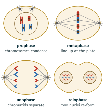
For convenience, nuclear division (karyokinesis) is divided into four stages, prophase, metaphase, anaphase and telophase; but cell division is a progressive process, and clear-cut lines cannot be drawn between the stages (page 122).
Prophase
Prophase, the first stage of karyokinesis, follows the S and G2 phases, in which the new DNA molecules are intertwined (page 122):
- Chromosomal material condenses into compact mitotic chromosomes, each seen to be made of two chromatids attached at the centromere.
- The centrosome was duplicated during interphase; the two centrosomes now begin to move towards opposite poles of the cell. Each radiates microtubules called asters; the two asters together with the spindle fibres form the mitotic apparatus. Centrosomes with centrioles are an animal-cell feature: almost all plant cells lack centrioles (NCERT Chapter 8, page 91), yet they still form a spindle.
- At the end of prophase, cells viewed under the microscope do not show the Golgi complexes, endoplasmic reticulum, nucleolus and nuclear envelope (page 123). They are rebuilt later: in telophase a nuclear envelope develops round each chromosome cluster, and the nucleolus, Golgi complex and ER reform (page 124). Beyond the NCERT text: in animal cells the nuclear envelope breaks up into small vesicles rather than vanishing (Wikipedia, “Mitosis”, facts in our words).
NCERT’s chapter Summary (page 129) puts it differently: “the centrioles move to the opposite poles” (in animal cells each centrosome carries a pair of centrioles; NCERT Chapter 8, page 99) and “The nuclear envelope and the nucleolus disappear and the spindle fibres start appearing.” So in an MCQ, the nuclear envelope disappears in prophase, by its end; its complete disintegration marks the start of metaphase (page 123). Beyond the NCERT text: accounts that add a stage called prometaphase between prophase and metaphase place the envelope’s breakdown there (Wikipedia, “Mitosis”, facts in our words).
Metaphase
- The complete disintegration of the nuclear envelope marks the start of metaphase, and the chromosomes spread through the cytoplasm. Condensation is complete, so this is the stage at which chromosome morphology is most easily studied (page 123).
- Each metaphase chromosome has two sister chromatids held together by the centromere. Small disc-shaped structures at the surface of the centromeres, the kinetochores, are the sites where spindle fibres attach (page 123).
- All the chromosomes come to lie at the equator, one chromatid of each connected by its kinetochore to spindle fibres from one pole and its sister chromatid to fibres from the opposite pole. This plane of alignment is the metaphase plate (page 123).
Anaphase
- At the start of anaphase, each chromosome at the metaphase plate splits simultaneously, and the two daughter chromatids, now called daughter chromosomes, begin moving towards opposite poles (page 123).
- The centromere of each chromosome stays directed towards the pole, at the leading edge, with the arms trailing behind (page 123).
- Key events: centromeres split and chromatids separate; chromatids move to opposite poles (page 124).
Telophase
- The chromosomes that have reached the poles decondense and lose their individuality; each set of chromatin collects at a pole (page 124).
- A nuclear envelope develops round each cluster, forming two daughter nuclei, and the nucleolus, Golgi complex and ER reform (page 124).
Cytokinesis
Cell division is completed when the cytoplasm divides, in cytokinesis (page 124). Beyond the NCERT text: cytokinesis typically starts once anaphase is under way, so it overlaps the last stages of nuclear division (Wikipedia, “Mitosis”, facts in our words). NCERT describes it as follows (page 124):
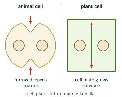
- Animal cells: a furrow appears in the plasma membrane, deepens and finally joins in the centre, dividing the cytoplasm into two.
- Plant cells: enclosed by a relatively inextensible cell wall, they divide differently. Wall formation starts in the centre and grows outward to meet the existing lateral walls. It begins with a simple precursor, the cell plate, which represents the middle lamella between the walls of the two cells.
- Organelles such as mitochondria and plastids are distributed between the daughter cells.
- In some organisms karyokinesis is not followed by cytokinesis, giving a multinucleate condition, a syncytium, as in the liquid endosperm of coconut (page 124). Beyond the NCERT text: a multinucleate cell formed by karyokinesis without cytokinesis is also called a coenocyte (Wikipedia, “Mitosis”, facts in our words); in an exam, use NCERT’s word, syncytium.
Solved examples
Example 1: Why “equational”? (NCERT Exercise 5)
Why is mitosis called equational division?
Answer: because the parent cell and the daughter cells have the same number of chromosomes (NCERT page 122): diploid cells give diploid cells, and the haploid cells that divide by mitosis, as in male honey bees, give haploid cells (NCERT page 122).
Example 2: Name the stage (NCERT Exercise 6, parts i and ii)
At which stage (i) are chromosomes moved to the spindle equator, and (ii) does the centromere split and the chromatids separate?
Answer: (i) metaphase; (ii) anaphase (NCERT pages 123–124). Centromeres also split in anaphase II of meiosis; parts (iii) and (iv) of this exercise, on pairing and crossing over, are answered on the meiosis page.
Example 3: Cytokinesis in plants and animals (NCERT Exercise 8)
How does cytokinesis in plant cells differ from that in animal cells?
Answer: an animal cell divides by a furrow in the plasma membrane that deepens from the outside inward; a plant cell, with an inextensible wall, forms a cell plate in the centre that grows outward to meet the lateral walls; the cell plate represents the middle lamella between the walls of the two cells (NCERT page 124).
Example 4: Counting chromatids
A cell with 8 chromosomes divides by mitosis. How many chromatids are at the metaphase plate, and how many daughter chromosomes reach each pole at anaphase?
Answer: 8 chromosomes × 2 chromatids = 16 chromatids at the plate; the 16 daughter chromosomes split 8 to each pole, so each daughter nucleus again has 8. For a short time in anaphase, before cytokinesis, the cell holds 16 chromosomes (NCERT pages 122–124).
Common mistakes to avoid
Common mistake
Saying homologous chromosomes separate in mitotic anaphase
In mitotic anaphase the centromeres split and the sister chromatids of each chromosome move to opposite poles (NCERT pages 123–124). Homologous chromosomes pair in meiosis, at zygotene of prophase I, and separate in anaphase I (NCERT pages 125–127); the meiosis page compares the two anaphases (NCERT Exercise 10).
Common mistake
Putting the cell plate at the edge of the cell
The cell plate starts in the centre and grows outward (NCERT page 124).
Common mistake
Choosing prophase for studying chromosome shape
Chromosome morphology is studied most easily at metaphase, when condensation is complete (NCERT page 123).
Quick revision summary
- Mitosis is the equational division: parent and daughter cells have the same chromosome number.
- Prophase: chromosomes condense, each made of two chromatids joined at the centromere; centrosomes move apart with asters; mitotic apparatus; Golgi, ER, nucleolus and envelope not seen by its end.
- Metaphase: envelope gone; kinetochores attach to spindle fibres; chromosomes on the metaphase plate; best stage to study chromosomes.
- Anaphase: centromeres split; daughter chromosomes move to the poles, centromere first.
- Telophase: chromosomes decondense; nuclear envelopes, nucleolus, Golgi and ER reform.
- Cytokinesis: furrow in animal cells, cell plate from the centre outward in plant cells; syncytium without cytokinesis (coconut endosperm).
Formula sheet
- mitosis: prophase, metaphase, anaphase, telophase; cytokinesis completes division
- metaphase: chromosomes on the metaphase plate
- anaphase: centromeres split, chromatids to opposite poles
- animal cytokinesis: furrow; plant cytokinesis: cell plate
- syncytium: karyokinesis without cytokinesis
Questions students ask
What is the difference between karyokinesis and cytokinesis?
Karyokinesis is the division of the nucleus, the separation of the daughter chromosomes into two daughter nuclei through prophase, metaphase, anaphase and telophase, while cytokinesis is the division of the cytoplasm that completes cell division: the M phase starts with karyokinesis and usually ends with cytokinesis (NCERT pages 121–124). Cytokinesis takes place by a furrow in animal cells and by a cell plate in plant cells (page 124). The two are separate events: in some organisms karyokinesis is not followed by cytokinesis, which leaves a multinucleate condition, a syncytium, as in the liquid endosperm of coconut (NCERT page 124).
What is the difference between telophase and cytokinesis?
Telophase is the last stage of karyokinesis, the nuclear division, and ends with a nuclear envelope round the chromosome cluster at each pole, while cytokinesis is the division of the cytoplasm that splits the cell into two daughter cells (NCERT page 124). Before cytokinesis is complete, two daughter nuclei can share one cytoplasm; cytokinesis completes their physical separation, sharing organelles such as mitochondria and plastids between them.
What is the difference between metaphase and anaphase?
In metaphase the chromosomes, each still two sister chromatids held together by the centromere, lie at the metaphase plate with the kinetochores of the sister chromatids attached to spindle fibres from opposite poles, while in anaphase the centromeres split and the sister chromatids, now called daughter chromosomes, move to opposite poles, centromere first (NCERT pages 123–124). So the number of chromosomes in the cell doubles at the start of anaphase: a human cell, with 46 chromosomes at metaphase (NCERT Chapter 8, page 101), has 92 daughter chromosomes in anaphase, 46 moving to each pole.
How many stages of mitosis are there?
Mitosis has four stages in NCERT: for convenience its nuclear division, karyokinesis, is divided into prophase, metaphase, anaphase and telophase, and cytokinesis then divides the cytoplasm (NCERT pages 122 and 124). Books that count five stages either add prometaphase, the stage between prophase and metaphase in which the nuclear envelope breaks down and the chromosomes attach to the spindle (beyond the NCERT text; Wikipedia, “Mitosis”, facts in our words), or count cytokinesis as a stage. NCERT places these events at the end of prophase and the start of metaphase, and stresses that very clear-cut lines cannot be drawn between the stages (NCERT pages 122–123).
Is the metaphase plate a real structure in the cell?
No, the metaphase plate is not a real structure: it is the plane of alignment of the chromosomes at metaphase, at the equator of the spindle (NCERT page 123). Do not confuse it with the cell plate, which is a real structure: the simple precursor of the new wall that a plant cell builds in its centre during cytokinesis, which represents the middle lamella between the walls of the two daughter cells (NCERT page 124).
Practice questions
Try each question first, then open the answer to check your working.
-
Q1. What do the two asters and the spindle fibres together form?
Show answerHide answer
The mitotic apparatus (NCERT page 122).
-
Q2. What are kinetochores?
Show answerHide answer
Small disc-shaped structures at the surface of the centromeres, where spindle fibres attach to the chromosomes (NCERT page 123).
-
Q3. Name the structures that re-form in telophase.
Show answerHide answer
A nuclear envelope round the chromosome cluster at each pole, giving two daughter nuclei, and the nucleolus, Golgi complex and ER (NCERT page 124).