Human excretory system and the nephron
NCERT Class 11 Biology Chapter 16 Section 16.1
Quick answer
The human urinary system comprises two kidneys, two ureters, a bladder and a urethra. Each kidney has about a million nephrons, which filter plasma and modify the filtrate before it enters collecting ducts. Long-looped juxtamedullary nephrons help establish the gradient needed for concentrated urine.
With your textbook
NCERT Biology (Class 11), Chapter 16 “Excretory Products and their Elimination”, Section 16.1 “Human Excretory System”, pages 206–208.
Open the official NCERT chapter (PDF, 1.8 MB, ncert.nic.in) (opens in a new tab)
Checked against the 2026-27 reprint on .
Human excretory system
Human excretory system
In humans, the excretory system consists of a pair of kidneys, one pair of ureters, a urinary bladder and a urethra (NCERT Section 16.1, page 206). The working unit of each kidney is the nephron:
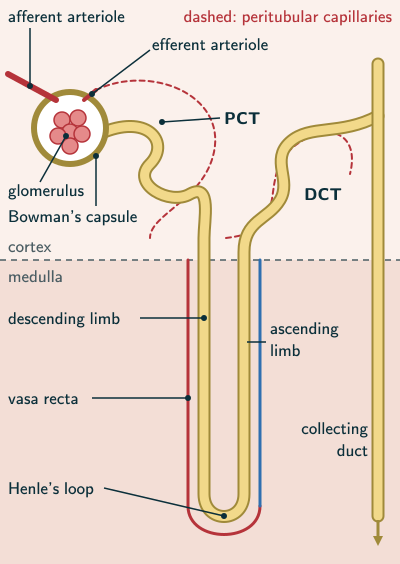
Each nephron has two parts – the glomerulus and the renal tubule.
NCERT Biology (Class 11), Section 16.1, page 207
In plain words: the two kidneys are the filters, and everything after them is plumbing. Inside each kidney about a million tiny tubes, the nephrons, each begin at a knot of capillaries that filters water and small solutes into a cup; the fluid runs down a long, looping tube, and what is left at the end drains into the funnel at the kidney’s centre and on to the bladder.
Kidneys, ureters, urinary bladder and urethra
NCERT describes the kidneys (page 206):
Kidneys are reddish brown, bean shaped structures situated between the levels of last thoracic and third lumbar vertebra close to the dorsal inner wall of the abdominal cavity.
NCERT Biology (Class 11), Section 16.1, page 206
- Each adult kidney is 10–12 cm long, 5–7 cm wide and 2–3 cm thick, with an average mass of 120–170 g (page 206).
- The urine formed in the nephrons is carried to the urinary bladder and stored there until it is released (NCERT Section 16.6, page 212).
- Beyond the NCERT text, the US National Institute of Diabetes and Digestive and Kidney Diseases (NIDDK) gives the role of each tube. The ureters are “Thin tubes of muscle that connect your kidneys to your bladder and carry urine to the bladder”, and the urethra is “A tube located at the bottom of the bladder that allows urine to exit the body during urination” (NIDDK, The Urinary Tract and How It Works).
- Compare the frog: it too has a pair of kidneys, ureters and a urinary bladder, which open into a cloaca (NCERT Chapter 7, pages 82–83).
Structure of the kidney
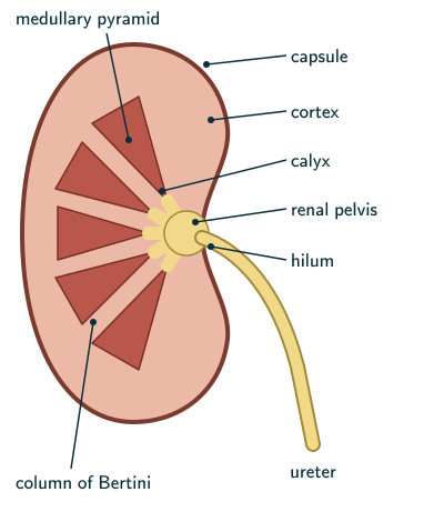
NCERT describes a kidney cut lengthwise (pages 206–207):
- Hilum: a notch towards the centre of the inner concave surface, through which blood vessels, nerves and the ureter pass. The renal artery enters, while the renal vein and ureter leave; NCERT’s word “enter” describes the hilum generally, not every direction of flow.
- Renal pelvis: a broad, funnel-shaped space inner to the hilum, with projections called calyces (singular: calyx).
- Capsule: the tough outer layer of the kidney.
- Cortex and medulla: inside are two zones, an outer cortex and an inner medulla.
- Medullary pyramids: the medulla is divided into a few conical masses that project into the calyces.
- Columns of Bertini: the cortex extends between the medullary pyramids as renal columns.
Nephron: glomerulus and renal tubule
- Each kidney has nearly one million complex tubular structures called nephrons, which are its functional units (NCERT page 207). NCERT’s own Summary says “over a million” (page 214); the two wordings differ slightly, but both round to about a million. NIDDK, beyond the NCERT text, also says each kidney “is made up of about a million filtering units called nephrons” (NIDDK, Your Kidneys and How They Work).
- Glomerulus: a tuft of capillaries formed by the afferent arteriole, a fine branch of the renal artery. Blood leaves the glomerulus by an efferent arteriole (page 207).
- Renal tubule (pages 207–208), in order:
- Bowman’s capsule: a double-walled, cup-like structure that encloses the glomerulus. The glomerulus with its Bowman’s capsule is the Malpighian body, or renal corpuscle.
- Proximal convoluted tubule (PCT): a highly coiled network.
- Henle’s loop: hairpin-shaped, with a descending and an ascending limb.
- Distal convoluted tubule (DCT): another highly coiled region.
- Collecting duct: not part of the nephron. The DCTs of many nephrons open into this straight tube, and many collecting ducts converge and open into the renal pelvis through the medullary pyramids in the calyces (page 208). NCERT’s Summary ends the renal tubule at the DCT (page 214).
- The PCT is lined by a simple cuboidal epithelium with a brush border (NCERT Section 16.3, page 209).
Cortical and juxtamedullary nephrons
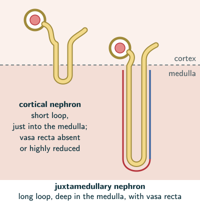
- The Malpighian corpuscle, PCT and DCT lie in the cortex; the loop of Henle dips into the medulla (NCERT page 208).
- Cortical nephrons: in the majority of nephrons, the loop of Henle is too short and extends only very little into the medulla (page 208).
- Juxtamedullary nephrons: in some nephrons, the loop of Henle is very long and runs deep into the medulla (page 208).
Peritubular capillaries and vasa recta
- The efferent arteriole leaving the glomerulus forms a fine capillary network around the renal tubule, the peritubular capillaries (NCERT page 208).
- A minute vessel of this network runs parallel to Henle’s loop, forming the U-shaped vasa recta (page 208).
- The vasa recta is absent or highly reduced in cortical nephrons (page 208).
Solved examples
Example 1: Name the parts (NCERT Exercise 11b, c)
Name (b) the cortical portions projecting between the medullary pyramids in the human kidney, and (c) a loop of capillary running parallel to Henle’s loop.
Answer: (b) the columns of Bertini (renal columns) (NCERT page 207); (c) the vasa recta (page 208).
Example 2: Follow the filtrate
Trace the path of fluid from the glomerulus to the renal pelvis.
Answer: glomerulus → Bowman’s capsule → PCT → descending limb of Henle’s loop → ascending limb → DCT → collecting duct → through the medullary pyramids into the calyces → renal pelvis (NCERT pages 207–208).
Example 3: How many nephrons?
Using NCERT’s figure, about how many nephrons are there in both kidneys together?
Answer: each kidney has nearly one million nephrons (NCERT page 207), so the two kidneys together have about 2 × 1 million = 2 million nephrons.
Example 4: Cortex or medulla?
Place each in the cortex or the medulla: (a) Malpighian corpuscle, (b) PCT, (c) the loop of a juxtamedullary nephron, (d) DCT, (e) medullary pyramids.
Answer: cortex: (a), (b) and (d); medulla: (c) and (e) (NCERT pages 206–208).
Example 5: The blood route
Name, in order, the vessels that carry blood into a glomerulus, out of it, and around the tubule.
Answer: the afferent arteriole (a fine branch of the renal artery) brings blood in; the efferent arteriole carries it out; it then forms the peritubular capillaries round the tubule, and the vasa recta beside Henle’s loop (NCERT pages 207–208).
Common mistakes to avoid
Common mistake
Calling Bowman’s capsule the renal corpuscle
The renal (Malpighian) corpuscle is the glomerulus together with its Bowman’s capsule; the capsule alone is the start of the renal tubule (NCERT page 207).
Common mistake
Giving every nephron a long loop
In the majority of nephrons, the cortical nephrons, the loop of Henle is short and extends only very little into the medulla; only the juxtamedullary nephrons have long loops deep in the medulla (NCERT page 208).
Common mistake
Counting the collecting duct as part of the nephron
A nephron ends at its DCT. The collecting duct is shared: the DCTs of many nephrons open into it (NCERT pages 208 and 214).
Common mistake
Putting the vasa recta in cortical nephrons
The vasa recta is absent or highly reduced in cortical nephrons (NCERT page 208).
Quick revision summary
- Excretory system: a pair of kidneys, a pair of ureters, a urinary bladder and a urethra. Kidneys lie between the last thoracic and third lumbar vertebrae; each is 10–12 cm long and 120–170 g.
- A kidney has a hilum, a renal pelvis with calyces, a tough capsule, an outer cortex and an inner medulla of pyramids, with columns of Bertini between them.
- Nearly one million nephrons per kidney; each is a glomerulus plus a renal tubule (Bowman’s capsule, PCT, Henle’s loop, DCT), opening into collecting ducts.
- Corpuscle, PCT and DCT are in the cortex; the loop dips into the medulla: short in cortical nephrons, long in juxtamedullary ones.
- The efferent arteriole forms peritubular capillaries and the U-shaped vasa recta, which is absent or highly reduced in cortical nephrons.
Formula sheet
- excretory system: 2 kidneys + 2 ureters + urinary bladder + urethra
- nephron = glomerulus + renal tubule
- renal corpuscle = glomerulus + Bowman’s capsule
- renal tubule: Bowman’s capsule → PCT → Henle’s loop → DCT; many DCTs → one collecting duct
- blood: afferent arteriole → glomerulus → efferent arteriole → peritubular capillaries, vasa recta
Questions students ask
Why are juxtamedullary nephrons important for making concentrated urine?
Juxtamedullary nephrons matter for concentrated urine because only they have very long loops of Henle running deep into the medulla, each with a vasa recta beside it; in cortical nephrons, the majority, the loop extends only very little into the medulla and the vasa recta is absent or highly reduced (NCERT page 208). The closeness of the loop and the vasa recta, and the counter currents in them, keep the medullary osmolarity rising from 300 mOsmol L in the cortex to about 1200 mOsmol L in the inner medulla, and, when ADH makes the collecting duct sufficiently water-permeable, that gradient draws water out of it (NCERT pages 211–212).
Why is the nephron called the functional unit of the kidney?
The nephron is called the functional unit of the kidney because each nephron performs filtration, reabsorption and secretion as part of an integrated kidney: NCERT calls the nearly one million nephrons in each kidney its functional units (page 207), and says glomerular filtration, reabsorption and secretion take place in different parts of the nephron (page 208). Its glomerulus filters plasma, and its tubule modifies the filtrate. The nephron tubules together with collecting ducts reabsorb nearly 99 per cent overall and secrete selected substances; shared blood vessels, the medullary gradient and hormones coordinate this work (NCERT pages 208–209).
How do you draw a labelled diagram of a nephron?
A labelled nephron, like NCERT Figure 16.3 (page 207), is drawn from the blood end to the duct: a glomerulus, the capillary tuft between an afferent and an efferent arteriole, inside a double-walled, cup-like Bowman’s capsule; then a coiled PCT, a hairpin Henle’s loop with descending and ascending limbs, and a coiled DCT opening into a straight collecting duct (NCERT pages 207–208). Draw the boundary of cortex and medulla, with the corpuscle, PCT and DCT above it and the loop dipping below (page 208). Bring the DCT back to touch the afferent arteriole, where the JGA forms (page 209), and add the peritubular capillaries, with the U-shaped vasa recta beside the loop.
Is the human excretory system in the NEET (UG) 2026 syllabus?
Yes, the human excretory system is in the NEET (UG) 2026 Biology syllabus: Unit 5, Human Physiology, lists “Human excretory system-structure and fuction” under “Excretory products and their elimination”, together with modes of excretion, urine formation, osmoregulation, regulation of kidney function, the role of other organs, disorders, and dialysis and the artificial kidney. The matching NCERT text is Class 11 Biology, Chapter 16: its opening pages and Sections 16.1 to 16.8 (pages 205–214), which this page and its eight sibling pages cover.
Practice questions
Try each question first, then open the answer to check your working.
-
Q1. What enters and leaves the kidney through the hilum?
Show answerHide answer
The renal artery enters; the renal vein and ureter leave. Nerves and lymphatics also pass through the hilum (NCERT page 206 lists the ureter, vessels and nerves).
-
Q2. What is a glomerulus?
Show answerHide answer
A tuft of capillaries formed by the afferent arteriole, a fine branch of the renal artery (NCERT page 207).
-
Q3. Which nephrons make up the majority, and how long are their loops?
Show answerHide answer
Cortical nephrons; their loops of Henle are too short and extend only very little into the medulla (NCERT page 208).