Genetic disorders
NCERT Class 12 Biology Chapter 4 Section 4.8
Quick answer
Genetic disorders can involve altered genes or chromosomes; new variants mean they are not always inherited from an affected parent. Pedigrees help infer inheritance, while examples include haemophilia, sickle-cell disease, PKU and thalassemia. Classic chromosome patterns include trisomy 21, 47, XXY and 45, X, with important mosaic, structural and clinical variation.
With your textbook
NCERT Biology (Class 12), Chapter 4 “Principles of Inheritance and Variation”, Section 4.8 “Genetic Disorders”, pages 72–76.
Open the official NCERT chapter (PDF, 19.2 MB, ncert.nic.in) (opens in a new tab)
Checked against the 2026-27 reprint on .
What are genetic disorders?
Genetic disorders
A number of disorders in human beings have been found to be associated with the inheritance of changed or altered genes or chromosomes.
NCERT Biology (Class 12), Section 4.8.1, page 73
- Mendelian disorders are mainly determined by alteration or mutation in a single gene (page 73).
- Chromosomal disorders are caused by the absence, excess or abnormal arrangement of one or more chromosomes (page 75).
In plain words: genetic disorders come from changed genes or chromosomes (NCERT page 73). Beyond the NCERT text, a genetic change may be inherited or arise newly, so “genetic” does not always mean inherited from an affected parent: most cases of Down’s, Klinefelter’s and Turner’s syndromes are not inherited (MedlinePlus Genetics, US National Library of Medicine, public domain). This lesson follows NCERT’s single-gene and chromosomal examples, which do not exhaust all genetic conditions.
Pedigree analysis
- The idea that disorders are inherited is old, based on characteristic features that run in families. Analysing the inheritance of traits in humans began after Mendel’s work was rediscovered (page 72).
- Controlled crosses, as in pea, are not possible in humans, so the study of a family’s history provides the alternative. An analysis of traits over several generations of a family is pedigree analysis: the trait is shown in a family tree (page 72).
- In human genetics, pedigree study is a strong tool to trace the inheritance of a specific trait, abnormality or disease (page 72).
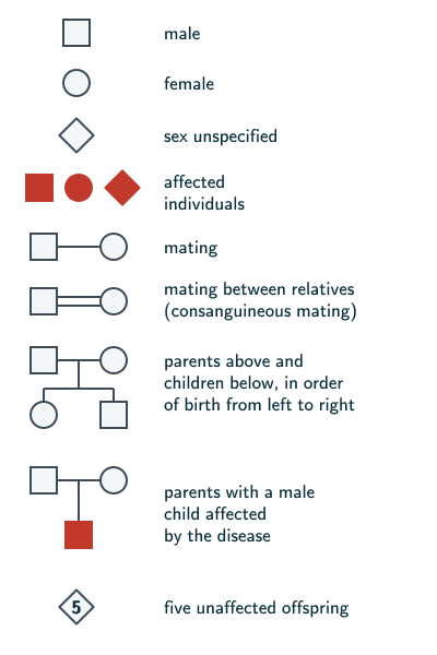
NCERT slip (page 72): NCERT numbers two figures 4.13: the honey bee (page 71) and these pedigree symbols (page 72). Cite the page with the figure number.
Mendelian disorders
- Mendelian disorders are transmitted to the offspring on the same lines as the principles of inheritance, and their pattern can be traced in a family by pedigree analysis (page 73).
- NCERT’s examples include haemophilia, cystic fibrosis, sickle-cell anaemia, colour blindness, phenylketonuria and thalassemia (page 73).
- They may be dominant or recessive, and pedigree analysis shows which. A trait may also be linked to the sex chromosome, as haemophilia is: this X-linked recessive trait passes from a carrier female to male progeny (page 73).
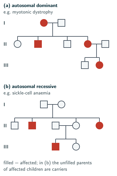
NCERT’s examples are myotonic dystrophy for an autosomal dominant trait and sickle-cell anaemia for an autosomal recessive one (page 73). How to read the pedigrees (our reading of NCERT Figure 4.14): in an ideal dominant pedigree with full penetrance and no new mutation, affected individuals have an affected parent. Recessive inheritance can allow unaffected carrier parents to have an affected child. Real pedigrees may be small or involve new variants, reduced penetrance or other complications; a pattern supports a model rather than proving a diagnosis.
For an X-linked recessive trait such as haemophilia (reasoned from NCERT pages 73–74): mostly males are affected; an affected son usually has an unaffected carrier mother; an affected father passes his X-linked allele to all daughters and no sons in the typical XX/XY model. Daughters are heterozygotes if their mother contributes a functioning allele. Beyond the NCERT text: a female carrier of haemophilia sometimes has some symptoms (MedlinePlus, Hemophilia; public domain).
Colour blindness
- A sex-linked recessive disorder due to a defect in either the red or green cone of the eye, resulting in failure to discriminate between red and green colour. The defect is due to mutation in certain genes on the X chromosome (page 73).
- It occurs in about 8 per cent of males and only about 0.4 per cent of females, because the genes are on the X chromosome: males have only one X, and females have two (page 73).
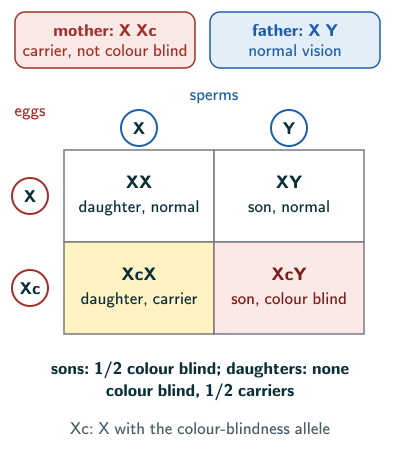
- The son of a woman who carries the gene has a 50 per cent chance of being colour blind. The mother is not colour blind herself, because the gene is recessive and her matching normal gene suppresses it. A daughter will not normally be colour blind, unless her mother is a carrier and her father colour blind (pages 73–74).
- Beyond the NCERT text: MedlinePlus Genetics describes red–green colour vision deficiency as trouble distinguishing between some shades of red and green. In its milder forms reds look less vivid, or greens look bluish or yellowish; only in the forms called protanopia and deuteranopia is red or green not perceived at all. It gives about 1 in 12 men and 1 in 200 women among people of Northern European ancestry; it is also common in East Asian populations and less common in almost all other populations studied. Other forms, such as blue–yellow deficiency, follow other inheritance patterns, and colour vision deficiency can also be acquired later in life.
Haemophilia
- A sex-linked recessive disease, transmitted from an unaffected carrier female to some of the male progeny (page 74).
- A single protein in the cascade of proteins involved in the clotting of blood is affected, so in an affected person a simple cut results in non-stop bleeding (page 74).
- The heterozygous (carrier) female may transmit the disease to her sons. A haemophilic female is extremely rare: her mother must be at least a carrier and her father haemophilic, a father NCERT describes as “unviable in the later stage of life” (page 74).
- The family pedigree of Queen Victoria shows a number of haemophilic descendants, as she was a carrier (page 74).
NCERT slip (page 74): NCERT calls the haemophilic father “unviable in the later stage of life”. That is too strong as a general statement: NCERT’s own route to a haemophilic daughter needs a haemophilic father who lives long enough to have children, and haemophilia is treated by replacing the missing clotting factor (beyond the NCERT text: MedlinePlus, Hemophilia; public domain). NCERT’s “non-stop bleeding” after a simple cut is also too strong: MedlinePlus describes “bleeding too much after an injury or surgery” and sudden bleeding inside the body, such as in the joints. In an exam, give NCERT’s reason why a haemophilic female is extremely rare: her mother must be at least a carrier and her father haemophilic.
Sickle-cell anaemia
- An autosome-linked recessive trait, transmitted when both partners are carriers (heterozygous). It is controlled by a single pair of alleles, HbA and HbS (page 74). NCERT names the case where “both the partners are carrier”, but it is not the only route: a parent with the disease (HbSHbS) passes on an HbS allele to every child, so a child of such a parent and a carrier has a 50 per cent chance of the disease (reasoned from NCERT page 74).
- Of the three possible genotypes, only HbSHbS shows the disease. HbAHbS people appear unaffected but are carriers, with a 50 per cent probability of transmitting the mutant gene to the progeny: they show the sickle-cell trait (page 74).
- The defect is the substitution of glutamic acid (Glu) by valine (Val) at the sixth position of the beta globin chain, from a single base substitution in the sixth codon. Under low oxygen tension the mutant haemoglobin polymerises, and the red blood cell changes from a biconcave disc to an elongated sickle-like structure (NCERT Figure 4.15, pages 74–75).
- NCERT slip (page 75): NCERT’s text gives the codon change as “from GAG to GUG”; that is the mRNA codon, and NCERT’s own Figure 4.15 shows the gene changing from GAG to GTG. In a written answer: gene GAG → GTG, mRNA GAG → GUG; specify that the DNA sequence is the coding strand read 5′→3′. In a multiple-choice question that repeats NCERT’s “GAG to GUG”, treat it as correct.
Scope (beyond the NCERT text): sickle-cell disease is a group of disorders that also includes HbSC and HbS/β-thalassemia; it is not limited to HbSS (MedlinePlus Genetics). People with sickle-cell trait are generally healthy, but they can pass the gene on to their children (MedlinePlus, Sickle Cell Disease; public domain).
Phenylketonuria
- An inborn error of metabolism, also inherited as an autosomal recessive trait (page 75).
- The affected individual lacks an enzyme that converts the amino acid phenylalanine into tyrosine. As a result, phenylalanine accumulates and is converted into phenylpyruvic acid and other derivatives; their accumulation in the brain results in mental retardation (NCERT’s term; now called intellectual disability), and they are also excreted through urine because of their poor absorption by the kidney (page 75). NCERT names the enzyme, phenyl alanine hydroxylase, on page 69; see pleiotropy.
Thalassemia
- Also an autosome-linked recessive blood disease, transmitted when both partners are unaffected carriers (heterozygous) (page 75).
- A mutation or deletion reduces the rate of synthesis of one of the globin chains (α or β) of haemoglobin. This causes the formation of abnormal haemoglobin molecules, resulting in the anaemia characteristic of the disease (page 75).
- Beyond the NCERT text: with too little haemoglobin, red blood cells do not develop normally (MedlinePlus Genetics, Beta thalassemia). In a small percentage of families a β thalassemia variant is inherited in an autosomal dominant manner, and some people with one altered HBB gene have mild anaemia, called thalassemia minor (MedlinePlus Genetics). Give NCERT’s autosomal recessive pattern, with unaffected carrier parents, in an NCERT-based answer.
- α thalassemia: production of the α globin chain is affected. It is controlled by two closely linked genes, HBA1 and HBA2, on chromosome 16; a person gets one chromosome 16 from each parent, so has four of these genes, and the disorder arises from mutation or deletion of one or more of the four; the more genes affected, the less α globin is made (page 75). Beyond the NCERT text: the kind of change also matters, as variants that alter the DNA sequence without deleting a gene tend to reduce α globin more than deletions (MedlinePlus Genetics, Alpha thalassemia).
- β thalassemia: production of the β globin chain is affected. It is controlled by a single gene, HBB, on chromosome 11, one copy from each parent, and arises from mutation of one or both copies (page 75).
- Thalassemia is a quantitative problem, too few globin molecules; sickle-cell anaemia is a qualitative problem, an incorrectly functioning globin (page 75).
Chromosomal disorders
Aneuploidy and polyploidy
- NCERT: “Failure of segregation of chromatids during cell division cycle results in the gain or loss of a chromosome(s), called aneuploidy”; examples are the extra chromosome 21 of Down’s syndrome and the loss of an X chromosome in Turner’s syndrome (page 75). Give this in an NCERT-based answer. Beyond the NCERT text: the failure can be of homologous chromosomes in meiosis I or of sister chromatids in meiosis II or mitosis (our reading of NCERT Class 11 Biology, Chapter 10).
- NCERT: failure of cytokinesis after the telophase stage of cell division results in an increase in a whole set of chromosomes in an organism, polyploidy, a condition often seen in plants (page 75). Give this in an NCERT-based answer. Beyond the NCERT text: polyploidy means more than two complete chromosome sets in a nucleus, and failed cytokinesis alone may at first leave two diploid nuclei rather than one polyploid nucleus (our reasoning).
- The total number of chromosomes in a normal human cell is 46 (23 pairs): 22 pairs of autosomes and one pair of sex chromosomes. An additional copy of a chromosome is a trisomy, and the lack of one of a pair a monosomy; such a situation leads to very serious consequences in the individual (pages 75–76). “Normal human cell” here means a nucleated diploid cell (reasoned from NCERT Section 2.3, page 31, where the secondary spermatocytes formed by reduction division “have only 23 chromosomes each”, and NCERT Class 11 Biology, Chapter 15, page 194: “RBCs are devoid of nucleus in most of the mammals”). The consequences differ with the chromosome, as the three syndromes below show (reasoned from NCERT page 76).
Down’s syndrome
- Cause: an additional copy of chromosome 21 (trisomy of 21); first described by Langdon Down (1866) (page 76).
- Features: short stature with a small round head, furrowed tongue and partially open mouth; a broad palm with a characteristic palm crease; physical, psychomotor and mental development is delayed (NCERT’s word is “retarded”) (page 76; NCERT Figure 4.16 also labels a flat back of the head, a broad flat face, a big and wrinkled tongue, many loops on the finger tips and congenital heart disease).
- Beyond the NCERT text: most cases are full trisomy 21, with 47 chromosomes. Less commonly, extra chromosome-21 material is attached to another chromosome (translocation Down’s syndrome), or the extra copy is in only some of the body’s cells (mosaic Down’s syndrome). Features vary from person to person; for example, about half of affected children are born with a heart defect (MedlinePlus Genetics).
Klinefelter’s syndrome
- Cause: an additional copy of the X chromosome, a karyotype of 47, XXY (page 76).
- Overall masculine development, but feminine development, such as breast development (gynaecomastia), is also expressed. Such individuals are sterile (page 76). Give this in an NCERT-based answer. NCERT Figure 4.17 (page 76) shows tall stature with feminised character.
- Beyond the NCERT text: features vary. Small testes make less testosterone, and infertility is common, but up to half of people with Klinefelter’s syndrome may be able to have children using assisted reproductive technologies; about 10 per cent have a mosaic form, with the extra X in only some cells (MedlinePlus Genetics).
Turner’s syndrome
- Cause: the absence of one of the X chromosomes, 45 with X0 (page 76). NCERT’s summary writes it XO (page 78); both mean one X and no second sex chromosome.
- Such females are sterile, as the ovaries are rudimentary, besides other features including lack of other secondary sexual characters (page 76). Give this in an NCERT-based answer. NCERT Figure 4.17 (page 76) shows short stature and underdeveloped feminine character.
- Beyond the NCERT text: the karyotype is also written 45,X. About half of people with Turner’s syndrome have one X in every cell; in others an X is partly missing or rearranged, or only some cells lack it (mosaic Turner’s syndrome). Short stature is the most common feature. Reduced ovarian function is very common and most are unable to become pregnant naturally, but a small percentage keep normal ovarian function through young adulthood (MedlinePlus Genetics).
Reading note: use respectful current terms such as intellectual disability and delayed development. Clinical descriptions are variable features, not labels that define every person with a condition.
Solved examples
Example 1: Pedigree analysis (NCERT Exercise 10)
What is pedigree analysis? Suggest how such an analysis can be useful.
Answer: pedigree analysis is the analysis of a trait over several generations of a family, shown as a family tree (NCERT page 72). It is useful because controlled crosses are not possible in humans: a pedigree traces the inheritance of a specific trait, abnormality or disease, helps infer whether a Mendelian disorder may be dominant, recessive or sex-linked, alongside clinical and genetic evidence (NCERT pages 72–73).
Example 2: Two autosomal disorders (NCERT Exercise 16)
Mention any two autosomal genetic disorders with their symptoms.
Answer:
- Sickle-cell anaemia (autosomal recessive, Mendelian): HbSHbS people have mutant haemoglobin that polymerises at low oxygen tension, so red blood cells change from biconcave discs to elongated sickle shapes (NCERT pages 74–75).
- Down’s syndrome (trisomy of chromosome 21, an autosome): short stature, small round head, furrowed tongue, partially open mouth, broad palm with a characteristic crease, delayed physical, psychomotor and mental development (NCERT page 76).
Phenylketonuria and thalassemia are other autosomal recessive examples (NCERT page 75).
Example 3: A carrier mother
A woman whose father was colour blind has normal vision. She marries a man with normal vision. What are the chances that (a) a son and (b) a daughter is colour blind?
Answer (simple fully recessive XX/XY model): she received her father’s only X, which carries the allele, so she is a carrier (X Xc). The father gives his X to every daughter and his Y to every son.
- (a) A son gets Xc or X from her, ½ each: ½ colour blind (NCERT page 74).
- (b) A daughter gets the father’s normal X, so she cannot be colour blind: 0; she is a carrier with chance ½.
Example 4: Two carriers of sickle-cell anaemia
Both parents are HbAHbS. What are the chances that a child (a) has the disease, (b) is a carrier?
Answer: each parent passes HbA or HbS, ½ each.
- (a) HbSHbS: ½ × ½ = ¼.
- (b) HbAHbS: ½ × ½ + ½ × ½ = ½.
Common mistakes to avoid
Common mistake
Calling sickle-cell anaemia sex-linked
NCERT’s sickle-cell/PKU/thalassemia examples are autosomal; its haemophilia and colour-blindness examples are X-linked recessive (NCERT pages 73–75).
Common mistake
Mixing up thalassemia and sickle-cell anaemia
Thalassemia is a quantitative problem, too few globin molecules; sickle-cell anaemia is a qualitative problem, an incorrectly functioning globin (NCERT page 75).
Common mistake
Swapping Klinefelter’s and Turner’s karyotypes
Klinefelter’s syndrome is 47, XXY (a male); Turner’s syndrome is 45, X0 (a female) (NCERT page 76).
Common mistake
Expecting a colour-blind son to get the allele from his father
A son gets his only X from his mother; the father passes his Y to sons (NCERT page 74).
Quick revision summary
- Pedigree analysis traces a trait over several generations of a family; controlled crosses are not possible in humans.
- Mendelian disorders come from a single gene: haemophilia, cystic fibrosis, sickle-cell anaemia, colour blindness, phenylketonuria, thalassemia.
- X-linked recessive: colour blindness (about 8 per cent of males, 0.4 per cent of females) and haemophilia; a carrier mother may pass haemophilia to her sons, and a haemophilic female is extremely rare.
- Autosomal recessive: sickle-cell anaemia (Glu → Val at position 6 of the beta chain), phenylketonuria, thalassemia (α: HBA1, HBA2 on chromosome 16; β: HBB on chromosome 11).
- Chromosomal disorders: aneuploidy (Down’s, trisomy 21; Turner’s, 45, X0; Klinefelter’s, 47, XXY) and polyploidy (failure of cytokinesis; extra complete sets). Beyond the NCERT text: translocation and mosaic forms also occur (MedlinePlus Genetics).
Formula sheet
- X-linked recessive: carrier mother × normal father → 1/2 of sons affected, daughters 1/2 carriers
- autosomal recessive: carrier × carrier → 1/4 affected, 1/2 carriers
- HbAHbS: carrier (sickle-cell trait); HbSHbS: diseased
- classic full trisomy 21: 47 chromosomes; translocation/mosaic Down’s syndrome also occurs
- Klinefelter’s syndrome: 47, XXY; Turner’s syndrome: 45, X0
Questions students ask
What is the difference between Mendelian and chromosomal disorders?
Mendelian disorders are mainly determined by alteration or mutation in a single gene, while chromosomal disorders are caused by the absence, excess or abnormal arrangement of one or more chromosomes (NCERT pages 73 and 75). A Mendelian disorder is transmitted on the same lines as Mendel’s principles, so its pattern can be traced through a family by pedigree analysis (page 73), while a chromosomal disorder alters the chromosome set itself. Sickle-cell anaemia and haemophilia are among NCERT’s Mendelian examples, and Down’s, Klinefelter’s and Turner’s syndromes are its chromosomal ones (pages 73 and 76).
Are chromosomal disorders inherited?
Beyond the NCERT text, most chromosomal disorders are not inherited, though they are genetic: MedlinePlus Genetics (US National Library of Medicine, 2021) explains that changes in chromosome number “are not inherited, but occur as random events during the formation of reproductive cells (eggs and sperm)”; NCERT calls such a change aneuploidy (page 75). Also beyond the NCERT text, some structural rearrangements can be passed on, as in translocation Down’s syndrome from an unaffected parent with a balanced rearrangement involving chromosome 21 (MedlinePlus Genetics, 2020).
Are all Mendelian disorders caused by point mutations?
No, Mendelian disorders are not all caused by point mutations: NCERT defines them as disorders “mainly determined by alteration or mutation in the single gene” (page 73), and that alteration can be larger than one base pair. Sickle-cell anaemia is NCERT’s classical point mutation, a single base substitution in the beta globin gene (pages 72 and 75), but thalassemia can arise from “either mutation or deletion”, and α thalassemia from the mutation or deletion of one or more of its four genes (page 75). What makes a disorder Mendelian is not the size of the change but its transmission on Mendel’s lines, which pedigree analysis can trace (page 73).
What is the difference between a pedigree chart and a Punnett square?
A pedigree chart records the traits that actually appeared in a family over several generations, while a Punnett square calculates the probability of each possible genotype among the offspring of one cross (NCERT pages 72 and 57). Used together, the pedigree reveals genotypes and the square turns them into chances: if an unaffected woman’s mother had sickle-cell anaemia, the woman must be HbAHbS, and a Punnett square of her alleles against her partner’s then gives the chance for each child (our working).
What is a karyotype, and which genetic disorders does it show?
Beyond the NCERT text, a karyotype is both a person’s complete set of chromosomes and the picture made when the chromosomes of one cell are photographed and lined up in numbered order, which shows whether any are missing, extra or rearranged (NHGRI Talking Glossary). NCERT’s summary says that Down’s, Turner’s and Klinefelter’s syndromes “can be easily studied by analysis of Karyotypes” (page 78). A Mendelian disorder such as sickle-cell anaemia leaves the karyotype normal, because one changed base pair alters neither the number nor the visible form of the chromosomes (our reading).
Practice questions
Try each question first, then open the answer to check your working.
-
Q1. Which genes control α thalassemia, and on which chromosome are they?
Show answerHide answer
Two closely linked genes, HBA1 and HBA2, on chromosome 16 of each parent (NCERT page 75).
-
Q2. Why is colour blindness much commoner in males than in females?
Show answerHide answer
Its genes are on the X chromosome and the trait is recessive; males have only one X, so one recessive allele makes a male colour blind, while a female needs it on both X chromosomes (NCERT page 73).
-
Q3. What is the karyotype in Turner’s syndrome, and why are such females sterile?
Show answerHide answer
45 with X0, from the absence of one X chromosome; such females are sterile because the ovaries are rudimentary (NCERT page 76). Beyond the NCERT text: a small percentage keep normal ovarian function through young adulthood (MedlinePlus Genetics).