The female reproductive system
NCERT Class 12 Biology Chapter 2 Section 2.2
Quick answer
The female reproductive system includes ovaries, oviducts, uterus, cervix, vagina and external genital structures, with associated mammary glands. Ovaries produce oocytes and hormones; oviducts receive the ovulated secondary oocyte and the uterus supports implantation. Milk is produced in alveoli and passes through branching ducts to the nipple.
With your textbook
NCERT Biology (Class 12), Chapter 2 “Human Reproduction”, Section 2.2 “The Female Reproductive System”, pages 28–31.
Open the official NCERT chapter (PDF, 64.5 MB, ncert.nic.in) (opens in a new tab)
Checked against the 2026-27 reprint on .
What is the female reproductive system?
The female reproductive system
- It consists of a pair of ovaries, a pair of oviducts, the uterus, cervix, vagina and the external genitalia, located in the pelvic region (page 28).
- Together with a pair of mammary glands, these parts are integrated structurally and functionally to support ovulation, fertilisation, pregnancy, birth and child care (page 28).
Ovaries are the primary female sex organs that produce the female gamete (ovum) and several steroid hormones (ovarian hormones).
NCERT Biology (Class 12), Section 2.2, page 28
In plain words: the ovaries make the eggs and the female hormones; the fimbriae help guide an ovulated secondary oocyte into an oviduct, which opens into the uterus; the cervix and vagina lead outwards; fertilisation, pregnancy and birth take place along this tract; the mammary glands feed the baby after birth.
The ovaries: cortex and medulla
- The ovaries lie one on each side of the lower abdomen. Each is about 2 to 4 cm long and is connected to the pelvic wall and the uterus by ligaments (page 28).
- Each ovary is covered by a thin epithelium, which encloses the ovarian stroma (page 28).
The stroma is divided into two zones – a peripheral cortex and an inner medulla.
NCERT Biology (Class 12), Section 2.2, page 28
The ovary as an endocrine gland is covered in Class 11. Ovarian follicles at different stages of development are embedded in the stroma (NCERT Summary, page 39); they are on the gametogenesis page.
Oviducts, uterus and vagina
The oviducts (fallopian tubes), uterus and vagina are the female accessory ducts (page 29).
Fallopian tube (pages 29–30): each is about 10–12 cm long and runs from near an ovary to the uterus. Its ovarian end opens into the peritoneal cavity; the tube is not directly continuous with the ovary. From the ovary end:
- Infundibulum: the funnel-shaped part closest to the ovary. Its edges carry finger-like projections, the fimbriae, which help collect the ovum after ovulation.
- Ampulla: the wider part that follows.
- Isthmus: the last part, with a narrow lumen; it joins the uterus.
Fertilisation takes place in the ampullary region of the tube (NCERT Section 2.5, page 35).
Uterus (page 30):
- It is single and is also called the womb; its shape is like an inverted pear. It is supported by ligaments attached to the pelvic wall. Beyond the NCERT text: the pelvic-floor muscles, with the surrounding tissues, also hold the pelvic organs, including the uterus, in place (NICHD, About Pelvic Floor Disorders).
- It opens into the vagina through a narrow cervix. The cavity of the cervix, the cervical canal, together with the vagina forms the birth canal.
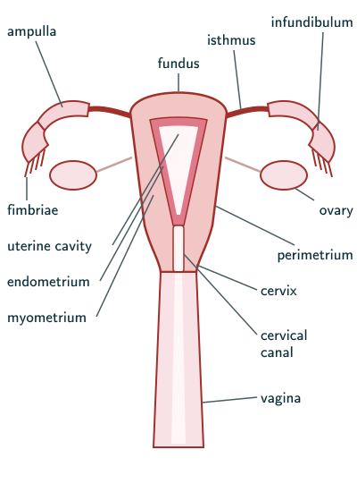
Layers of the uterine wall
The wall of the uterus has three layers of tissue (page 30):
- Perimetrium: the external, thin, membranous layer.
- Myometrium: the middle, thick layer of smooth muscle.
- Endometrium: the inner, glandular layer that lines the uterine cavity.
The endometrium undergoes cyclical changes during menstrual cycle while the myometrium exhibits strong contraction during delivery of the baby.
NCERT Biology (Class 12), Section 2.2, page 30
Female external genitalia
The female external genitalia are the mons pubis, labia majora, labia minora, hymen and clitoris (page 30):
- Mons pubis: a cushion of fatty tissue covered by skin and pubic hair.
- Labia majora: fleshy folds of tissue that extend down from the mons pubis and surround the vaginal opening.
- Labia minora: paired folds of tissue under the labia majora.
- Hymen: a membrane that often partially covers the opening of the vagina.
- Clitoris: its visible glans lies near the upper junction of the labia minora, above the urethral opening. Most of the organ, including its body and crura, is internal; NCERT’s small external structure describes only the visible part (NCBI Bookshelf, How do the female sex organs work?).
Hymenal tissue varies naturally in shape and elasticity. It may stretch without tearing, and its appearance cannot establish whether intercourse has occurred. WHO states that no examination can prove a history of vaginal intercourse (Eliminating virginity testing, 2018). Thus an “intact/broken” model should not be used as a test of sexual history.
In fact, the presence or absence of hymen is not a reliable indicator of virginity or sexual experience.
NCERT Biology (Class 12), Section 2.2, page 30
Mammary glands
A functional mammary gland is characteristic of all female mammals.
NCERT Biology (Class 12), Section 2.2, page 31
- The mammary glands are paired structures (breasts) with glandular tissue and a variable amount of fat (page 31).
- The glandular tissue of each breast is divided into 15–20 mammary lobes, which contain clusters of cells called alveoli. The cells of the alveoli secrete milk, which is stored in the cavities (lumens) of the alveoli (page 31).
- NCERT’s schematic duct sequence (page 31): alveoli → mammary tubules (the tubules of each lobe join) → mammary duct (several ducts join) → mammary ampulla → lactiferous duct → nipple, from where milk is expressed during breastfeeding.
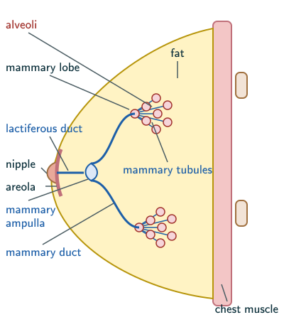
Modern anatomical qualification: ultrasound imaging found branching milk ducts without the large, typical lactiferous sinuses depicted in older diagrams (Ramsay and colleagues, Journal of Anatomy, 2005). Milk is produced in alveoli and conveyed through ducts to nipple openings; do not assume a fixed collecting ampulla in every breast.
Solved examples
Example 1: Two functions of the ovary (NCERT Exercise 4, second part)
Write two major functions of the ovary.
Answer: it produces the female gamete, the ovum, and it secretes several steroid hormones, the ovarian hormones (NCERT page 28).
Example 2: Endometrium and fimbriae (NCERT Exercise 15 b and e)
Name the functions of (a) the endometrium and (b) the fimbriae.
Answer: (a) the endometrium, the inner glandular layer of the uterus, lines the uterine cavity and undergoes cyclical changes during the menstrual cycle (NCERT page 30); it is the layer in which the blastocyst implants, and progesterone maintains it for pregnancy (NCERT pages 35 and 37). (b) The fimbriae, finger-like projections at the edge of the infundibulum, help collect the ovum after ovulation (NCERT page 29).
Example 3: From the ovary to the outside
List, in order, the parts of the female reproductive tract from the ovary to the outside of the body.
Answer: fimbriae and infundibulum → ampulla → isthmus → uterine cavity → cervix (cervical canal) → vagina (NCERT pages 29–30). The cervical canal and the vagina together form the birth canal (page 30).
Common mistakes to avoid
Common mistake
Putting the isthmus next to the ovary
The infundibulum, with its fimbriae, is nearest the ovary; then the ampulla; the narrow isthmus joins the uterus (NCERT pages 29–30).
Common mistake
Swapping endometrium and perimetrium
The endometrium is the inner glandular lining that changes in each cycle; the perimetrium is the thin outer membrane; the myometrium in between contracts at delivery (NCERT page 30).
Common mistake
Calling the ovaries accessory organs
Ovaries are the primary female sex organs; the oviducts, uterus and vagina are the accessory ducts (NCERT pages 28–29).
Quick revision summary
- Female system: two ovaries, two oviducts, uterus, cervix, vagina, external genitalia and two mammary glands, for ovulation, fertilisation, pregnancy, birth and child care.
- Ovaries (2 to 4 cm): primary sex organs making ova and steroid hormones; stroma with an outer cortex and an inner medulla.
- Fallopian tube (10–12 cm): infundibulum with fimbriae, ampulla, isthmus; then the inverted-pear uterus, the cervix and the vagina; cervical canal and vagina form the birth canal.
- Uterine wall: perimetrium, myometrium (contracts at delivery), endometrium (changes in each cycle).
- External genitalia: mons pubis, labia majora, labia minora, hymen, clitoris; the hymen is not a reliable indicator of virginity.
- Mammary gland: NCERT describes 15–20 lobes of alveoli and a tubule–duct–ampulla sequence. Modern imaging qualifies the ampulla model; milk travels from alveoli through branching ducts to the nipple.
Formula sheet
- ovary: 2 to 4 cm; cortex outside, medulla inside
- fallopian tube: 10–12 cm; fimbriae at the infundibulum; infundibulum → ampulla → isthmus → uterus
- uterine wall: perimetrium, myometrium, endometrium
- birth canal = cervical canal + vagina
- milk: alveoli → branching ducts → nipple (NCERT additionally labels an ampulla)
Questions students ask
Are the oviduct, fallopian tube and uterine tube the same?
Yes, in the human female the oviduct, the fallopian tube and the uterine tube are three names for the same paired tube: NCERT writes “oviducts (fallopian tubes)” (NCERT page 29), and beyond the NCERT text, MeSH lists oviducts and uterine tubes among the names of the fallopian tubes (MeSH, Fallopian Tubes). Each is about 10–12 cm long and extends from the periphery of an ovary to the uterus (page 29). NCERT also uses the word oviduct for other animals, such as the frog, whose pair of oviducts arises from the ovaries and opens into the cloaca (frog, NCERT Class 11, page 84).
What is the difference between testis and ovary?
The testis and the ovary are the primary sex organs that produce the gametes (NCERT page 31), but they differ in place, size, structure and timing. The testes lie in the scrotum outside the abdominal cavity, each about 4 to 5 cm long; the ovaries lie one on each side of the lower abdomen, each about 2 to 4 cm long (pages 27–28). A testis has about 250 lobules of seminiferous tubules; an ovary has a stroma of outer cortex and inner medulla holding follicles at different stages (pages 27–28 and 39). Spermatogenesis starts at puberty and “sperm formation continues even in old men”, but oogenesis starts before birth and menstrual cycles cease around 50 years of age (pages 26, 31, 32 and 35).
Is the mammary gland an exocrine or an endocrine gland?
The mammary gland is an exocrine gland: the milk secreted by the cells of its alveoli leaves through a branching system of tubules and ducts that opens at the nipple (NCERT page 31). Beyond the NCERT text, the NCI SEER training module names the mammary glands as exocrine glands, which “have ducts that carry their secretory product to a surface” (NCI SEER Training Modules, Introduction to the Endocrine System). Endocrine glands work the other way: they lack ducts, which is why NCERT calls them ductless glands (endocrine glands, NCERT Class 11, page 239).
Are the mammary glands and the breasts the same?
In NCERT the mammary glands are the breasts: it describes them as “paired structures (breasts) that contain glandular tissue and variable amount of fat” (NCERT page 31). Beyond the NCERT text, MeSH uses the two words differently: mammary gland for the milk-secreting tissue alone, and breast for the whole organ in which that tissue sits together with fat, skin, muscle and connective tissue (MeSH, Breast; Mammary Glands, Human). In each breast the glandular tissue is divided into 15–20 mammary lobes, whose alveoli secrete the milk (page 31).
Why are the fallopian tubes important?
The fallopian tubes are important because the ovum, the sperms and the early embryo all pass through them: each tube collects the ovum after ovulation (NCERT page 29) and carries it to its ampullary region, where fertilisation takes place (insemination, page 35), and the zygote starts cleavage as it moves through the isthmus towards the uterus (page 36). So the zygote forms in the tube, but pregnancy begins only when the blastocyst implants in the uterus (implantation, page 37).
Practice questions
Try each question first, then open the answer to check your working.
-
Q1. Which layer of the uterine wall shows strong contractions during the delivery of the baby?
Show answerHide answer
The myometrium, the middle layer of smooth muscle (NCERT page 30).
-
Q2. What is the funnel-shaped part of the fallopian tube nearest the ovary called, and what do its edges carry?
Show answerHide answer
The infundibulum; its edges carry finger-like fimbriae, which help collect the ovum after ovulation (NCERT page 29).
-
Q3. True or false: the presence or absence of the hymen is not a reliable indicator of virginity or sexual experience. (NCERT Exercise 16 g)
Show answerHide answer
True (NCERT page 30).