Respiratory organs and the human respiratory system
NCERT Class 11 Biology Chapter 14 Section 14.1
Quick answer
Air travels through the nose, pharynx, larynx, trachea and branching airways to the alveoli. The conducting region ends at terminal bronchioles; the alveolated respiratory region exchanges gases with blood. Pleura couple the lungs to chest-wall movement, while other animals may use body surfaces, tracheal tubes, gills or lungs.
With your textbook
NCERT Biology (Class 11), Chapter 14 “Breathing and Exchange of Gases”, Section 14.1 “Respiratory Organs”, pages 183–185.
Open the official NCERT chapter (PDF, 2.1 MB, ncert.nic.in) (opens in a new tab)
Checked against the 2026-27 reprint on .
Breathing and respiratory organs
Breathing
Most human cells use oxygen, indirectly, to break down simple molecules such as glucose, amino acids and fatty acids for energy, and they release carbon dioxide, whose excessive accumulation is harmful. So O must be supplied to the cells continuously and CO removed (NCERT page 183).
This process of exchange of O from the atmosphere with CO produced by the cells is called breathing, commonly known as respiration.
NCERT Biology (Class 11), Chapter 14, Introduction, page 183
In plain words: most body tissues need a steady oxygen supply and release carbon dioxide during metabolism. Mature human RBCs are an exception: they carry oxygen but lack mitochondria and do not consume it in aerobic respiration. Breathing is the delivery and waste-collection service: the lungs bring air close to the blood, and the blood carries the gases between the lungs and every cell.
Respiratory organs in animals
The mechanism of breathing varies among animal groups, depending mainly on their habitat and level of organisation (NCERT Section 14.1, page 183):
| Animals | Respiratory surface or organ |
|---|---|
| sponges, coelenterates, flatworms | entire body surface, by simple diffusion |
| earthworms | moist cuticle |
| insects | network of tracheal tubes carrying air in the body |
| most aquatic arthropods and molluscs | gills (branchial respiration) |
| terrestrial forms in NCERT’s comparison | lungs (pulmonary respiration) |
| fishes | gills |
| amphibians, reptiles, birds, mammals | lungs |
| frogs (also) | moist skin (cutaneous respiration) |
This table gives NCERT’s broad patterns, not rules without exceptions; for example, it already lists terrestrial insects using tracheae.
Gills are special vascularised structures, and lungs are vascularised bags (page 183). See the frog’s cutaneous and pulmonary respiration.
Human respiratory system
The air passes through these parts in order (NCERT Section 14.1.1, pages 184–185):
- A pair of external nostrils, opening above the upper lip, leads through the nasal passage into the nasal chamber.
- The nasal chamber opens into the pharynx, part of which is the common passage for food and air.
- The pharynx opens through the larynx into the trachea. The larynx is a cartilaginous box that helps in sound production, so it is called the sound box. During swallowing, the glottis can be covered by a thin, elastic, cartilaginous flap, the epiglottis, which stops food entering the larynx.
- The trachea is a straight tube running to the mid-thoracic cavity, where it divides, at the 5th thoracic vertebra in NCERT’s account, into the right and left primary bronchi.
- Each bronchus divides repeatedly into secondary and tertiary bronchi and bronchioles, ending in very thin terminal bronchioles. NCERT includes the trachea, bronchi and “initial bronchioles” under incomplete cartilaginous rings. Modern histology distinguishes them: tracheal rings give way to bronchial cartilage plates, and bronchioles lack cartilage (StatPearls, “Histology, Lung” and “Histology, Respiratory Epithelium”).
- Beyond NCERT’s shortened sequence, terminal bronchioles lead to respiratory bronchioles, alveolar ducts and alveolar sacs containing thin-walled alveoli. Respiratory bronchioles already have alveoli in their walls (StatPearls, “Histology, Lung”).
Modern anatomical descriptions place the carina around T4–T5 rather than treating T5 as an invariant level (StatPearls, “Anatomy, Head and Neck, Trachea”).
The branching network of bronchi, bronchioles and alveoli makes up the lungs. The two lungs are covered by a double-layered pleura, with pleural fluid between the layers, which reduces friction on the lung surface. The outer pleural membrane is in close contact with the thoracic lining, and the inner one with the lung surface (pages 184–185).
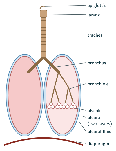
The lungs sit in the thoracic chamber, an anatomically air-tight chamber formed dorsally by the vertebral column, ventrally by the sternum, laterally by the ribs and below by the dome-shaped diaphragm. Normally, pleural coupling transmits chest-wall movement to the lungs, changing their volume. The airway remains open to the outside; “air-tight” describes the intact surrounding chest/pleural arrangement, not a sealed airway. This arrangement is essential for breathing, because we cannot alter the pulmonary volume directly (NCERT page 185).
Conducting and respiratory parts
The part starting with the external nostrils up to the terminal bronchioles constitute the conducting part whereas the alveoli and their ducts form the respiratory or exchange part of the respiratory system.
NCERT Biology (Class 11), Section 14.1.1, page 185
- The conducting part transports atmospheric air to the alveoli, clears it of foreign particles, humidifies it and brings it to body temperature (page 185).
- The exchange part is the site of diffusion of O and CO between blood and alveolar air (page 185). NCERT’s quoted list is abbreviated; modern anatomy includes respiratory bronchioles with alveoli as well as alveolar ducts, sacs and alveoli.
Steps of respiration
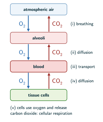
NCERT lists five steps (page 185):
- Breathing or pulmonary ventilation: atmospheric air is drawn in and CO-rich alveolar air is released out.
- Diffusion of O and CO across the alveolar membrane.
- Transport of gases by the blood.
- Diffusion of O and CO between blood and tissues.
- Use of O by the cells for catabolic reactions, with the release of CO: cellular respiration, dealt with in Chapter 12.
Solved examples
Example 1: Match the animal to its respiratory organ
Name the respiratory surface or organ in (a) a sponge, (b) an earthworm, (c) a cockroach, (d) a fish, (e) a frog in water.
Answer: (a) the entire body surface, by simple diffusion; (b) its moist cuticle; (c) a network of tracheal tubes; (d) gills; (e) its moist skin, cutaneous respiration (NCERT page 183; Chapter 7, page 82).
Example 2: Gas exchange in insects (NCERT Exercise 10)
What is the site of gaseous exchange in an insect?
Answer: the fine terminal branches of the tracheal system, the tracheoles, bring air close to tissue cells. Insects have a network of tubes, the tracheal tubes, that carry atmospheric air within the body (NCERT page 183).
Example 3: Only in the alveoli (NCERT Exercise 3)
Diffusion of gases occurs in the alveolar region only and not in the other parts of the respiratory system. Why?
Answer: the part from the external nostrils to the terminal bronchioles is only the conducting part: it transports, cleans and humidifies the air and brings it to body temperature. The alveolated respiratory region has thin walls, extensive surface area and nearby capillaries for diffusion; respiratory bronchioles also belong to this region in modern anatomy (NCERT pages 184–185).
Example 4: The path of a breath
List, in order, the parts air passes through from the nostrils to the alveoli.
Answer: external nostrils → nasal passage → nasal chamber → pharynx → larynx → trachea → primary bronchi → secondary and tertiary bronchi → bronchioles → terminal bronchioles → respiratory bronchioles → alveolar ducts/sacs → alveoli (NCERT’s page 184 sequence expanded using StatPearls, “Histology, Lung”).
Example 5: Why an air-tight thorax?
Why must the thoracic chamber be air-tight for breathing to work?
Answer: we cannot change the volume of the lungs directly. With intact pleural coupling, expansion of the chest wall lowers pleural pressure and expands the lungs, so moving the diaphragm and ribs changes the lung volume (NCERT page 185).
Common mistakes to avoid
Common mistake
Equating breathing with cellular respiration
NCERT uses “breathing” in two senses. On page 183 it is the whole exchange of atmospheric O with the CO made by cells, “commonly known as respiration”. In the list of steps on page 185 it is only step (i), pulmonary ventilation, which moves air between the atmosphere and the alveoli. Neither is cellular respiration, the use of O inside cells for catabolic reactions, which is step (v) (NCERT pages 183 and 185).
Common mistake
Putting gas exchange in the bronchi
From the nostrils to the terminal bronchioles is the conducting part (NCERT page 185). The alveolated respiratory region exchanges gases; modern anatomy includes respiratory bronchioles.
Common mistake
Getting the epiglottis wrong
The epiglottis is a flap that covers the glottis during swallowing, stopping food entering the larynx; the larynx itself is the sound box (NCERT page 184).
Quick revision summary
- Breathing is the exchange of atmospheric O with the CO produced by cells. Lower invertebrates use the body surface, earthworms the moist cuticle, insects tracheal tubes, most aquatic arthropods and molluscs and typical fishes gills, and typical adult land vertebrates lungs; frogs also use their skin.
- Human airway: nostrils → nasal chamber → pharynx → larynx (sound box; epiglottis) → trachea → bronchi (at the 5th thoracic vertebra) → bronchioles → alveoli.
- Lungs are covered by a double-layered pleura with pleural fluid, inside an air-tight thoracic chamber closed below by the diaphragm.
- Conducting part: nostrils to terminal bronchioles (transport, clean, humidify, bring to body temperature). Exchange part: the alveolated respiratory region, including respiratory bronchioles in modern anatomy.
- Steps: breathing (pulmonary ventilation), diffusion at the alveoli, transport by blood, diffusion at the tissues, use of O by cells.
Formula sheet
- airway: nostrils → nasal chamber → pharynx → larynx → trachea → bronchi → bronchioles → alveoli
- conducting part: nostrils to terminal bronchioles; exchange part: alveolated respiratory region
- NCERT: trachea divides at the 5th thoracic vertebra
- steps: breathing → alveolar diffusion → transport → tissue diffusion → cellular respiration
Questions students ask
What is the difference between breathing and respiration?
Breathing is the physical movement of air into and out of the lungs, while cellular respiration is the chemical oxidation of food inside the cells that releases energy. Breathing, or pulmonary ventilation, is done by muscles that change the volume of the thorax, about 12–16 times a minute in a healthy human (NCERT pages 185–186); cellular respiration goes on in the cytoplasm and mitochondria and traps the energy as ATP (NCERT Chapter 12, page 154). The two are linked: breathing supplies the O that the cells use and removes the CO they make (NCERT page 183).
What is the difference between terminal bronchioles and respiratory bronchioles?
Terminal bronchioles are the last tubes of the conducting part and carry no alveoli, while respiratory bronchioles, which NCERT does not name, are the narrower branches after them whose walls already carry some alveoli and so take part in gas exchange. NCERT ends the conducting part at the terminal bronchioles and says each terminal bronchiole gives rise to a number of alveoli (NCERT pages 184–185). Beyond the NCERT text, terminal bronchioles lead into several orders of respiratory bronchioles, then alveolar ducts and alveoli (MeSH, US National Library of Medicine, “Bronchioles”; StatPearls, “Histology, Lung”). The conducting/respiratory boundary is still at the end of the terminal bronchioles; the modern description fills in structures NCERT omits.
How is a frog’s respiration different from a human’s?
A frog uses more than one respiratory surface, while a human relies on its lungs: in water a frog respires through its skin (cutaneous respiration), and on land its buccal cavity, skin and lungs all act as respiratory organs (NCERT Chapter 7, page 82). During aestivation and hibernation the frog exchanges gases through the skin (page 82), whereas in humans the lungs alone serve, sitting in an air-tight thorax closed below by the diaphragm (NCERT pages 184–185). NCERT Section 7.2.2 on the frog covers its cutaneous and pulmonary respiration, including its lungs (page 82).
Why are the cartilaginous rings of the trachea incomplete?
The trachea’s cartilaginous rings are incomplete so that they hold the windpipe open while leaving a soft back wall next to the oesophagus, a reason NCERT does not give: it describes them only as incomplete cartilaginous rings that support the airway (NCERT page 184). Beyond the NCERT text, the trachea has 16 to 20 C-shaped rings of hyaline cartilage, open at the back, that stop it collapsing during inspiration. Their open ends are bridged by a smooth muscle, the trachealis, so the back wall gives way as the oesophagus expands during swallowing, and the muscle narrows the airway during coughing (StatPearls, “Anatomy, Head and Neck, Trachea”).
Practice questions
Try each question first, then open the answer to check your working.
-
Q1. What is the function of the pleural fluid?
Show answerHide answer
It reduces friction on the lung surface (NCERT page 184).
-
Q2. Which structure is called the sound box?
Show answerHide answer
The larynx, a cartilaginous box that helps in sound production (NCERT page 184).
-
Q3. Name the four structures that form the walls of the thoracic chamber.
Show answerHide answer
The vertebral column (dorsally), the sternum (ventrally), the ribs (laterally) and the dome-shaped diaphragm (below) (NCERT page 185).