Heart, cardiac cycle and ECG
NCERT Class 11 Biology Chapter 15 Section 15.3
Quick answer
Humans have a four-chambered heart, blood and a closed network of vessels. The sino-atrial node starts the beat; valves maintain one-way flow, and cardiac output equals stroke volume times heart rate. An ECG records electrical activity through P waves, QRS complexes and T waves.
With your textbook
NCERT Biology (Class 11), Chapter 15 “Body Fluids and Circulation”, Section 15.3 “Circulatory Pathways”, pages 197–201.
Open the official NCERT chapter (PDF, 3.1 MB, ncert.nic.in) (opens in a new tab)
Checked against the 2026-27 reprint on .
Circulatory pathways
Circulatory pathways
The circulatory patterns of animals are of two types, open and closed (NCERT Section 15.3, page 197). In humans, the circulatory system, also called the blood vascular system, has three parts:
Human circulatory system, also called the blood vascular system consists of a muscular chambered heart, a network of closed branching blood vessels and blood, the fluid which is circulated.
NCERT Biology (Class 11), Section 15.3.1, page 198
In plain words: the heart is a pump with four rooms and one-way doors. A tiny built-in timer in its wall fires about 72 times a minute; each beat squeezes first the upper rooms, then the lower ones, sending blood out through the arteries, and an ECG records the heart’s electrical activity from the skin.
Open and closed circulation
| Feature | Open | Closed |
|---|---|---|
| Found in | arthropods, molluscs | annelids, chordates |
| Path of blood | vessels, then open sinuses | always in vessels |
| Control of flow | less precise | more precise |
(NCERT page 197.) In an open system, blood pumped by the heart passes through large vessels into open spaces or body cavities called sinuses. In a closed system, the blood is always circulated through a closed network of blood vessels; this is considered more advantageous because the flow can be more precisely regulated (page 197). Beyond the NCERT text: cephalopod molluscs (octopuses, squid) have a closed system (see Common mistakes below).
Hearts of vertebrates
All vertebrates have a muscular, chambered heart (NCERT page 197):
- Fishes: a 2-chambered heart, one atrium and one ventricle. It pumps out deoxygenated blood, which is oxygenated by the gills and supplied to the body, and deoxygenated blood returns to the heart: single circulation.
- Amphibians and reptiles (except crocodiles): a 3-chambered heart, two atria and one ventricle. The left atrium receives oxygenated blood (from gills, lungs or skin) and the right atrium deoxygenated blood, but they mix in the single ventricle, which pumps out mixed blood: incomplete double circulation.
- Crocodiles, birds and mammals: a 4-chambered heart, two atria and two ventricles.
- Birds and mammals: oxygenated and deoxygenated blood pass from the left and right atria to the ventricles of the same sides and are pumped out without mixing: double circulation. NCERT names only birds and mammals here, not crocodiles.
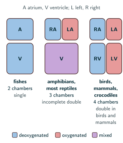
NCERT’s chamber-count scheme is a useful exam summary, but one ventricle does not imply complete mixing. Partial septa and flow patterns can separate streams; pythons and monitor lizards achieve strong functional separation of pulmonary and systemic flow and pressure (Jensen and colleagues, “How the python heart separates pulmonary and systemic blood pressures and blood flows”, Journal of Experimental Biology, 2010).
Human circulatory system
- The heart is mesodermally derived, lies in the thoracic cavity between the two lungs, slightly tilted to the left, and is the size of a clenched fist. It is protected by a double-walled membranous bag, the pericardium, enclosing pericardial fluid (NCERT page 198).
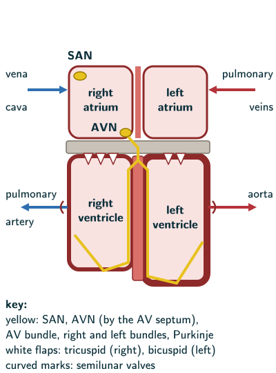
- It has four chambers: two relatively small upper atria and two larger lower ventricles (page 198).
- A thin, muscular inter-atrial septum separates the right and left atria; a thick-walled inter-ventricular septum separates the ventricles; the atrium and ventricle of the same side are separated by a thick fibrous atrio-ventricular septum (page 198). NCERT says “each of these septa” has an opening, but the openings join the two chambers of the same side, so they are in the atrio-ventricular septum, one on each side, guarded by the valves below (page 198).
- Valves (pages 198–199):
- tricuspid valve (three flaps or cusps): between the right atrium and the right ventricle. Beyond the NCERT text, the valve flaps are held by cords, the chordae tendineae, attached to papillary muscles; they stop the flaps from being pushed back into the atria when the ventricles contract (Ross and colleagues, Bioengineering, 2020);
- bicuspid (mitral) valve: between the left atrium and the left ventricle;
- semilunar valves: at the openings of the right ventricle into the pulmonary artery and of the left ventricle into the aorta.
- The valves let blood flow only one way, from atria to ventricles and from ventricles to the pulmonary artery or aorta, preventing backflow (page 199).
- Cardiac muscle forms the myocardium, the muscular part of the heart wall; the heart also contains connective tissue, an endothelial lining, vessels and nerves. The ventricle walls are much thicker than the atrial walls (NCERT page 199). NCERT calls the tricuspid cusps “muscular flaps”, but valve leaflets are connective tissue covered by endothelium, supported by chordae and papillary muscles (Charles University, Cardiovascular system anatomy notes). See cardiac muscle.
Nodal tissue and the pacemaker
A specialised cardiac musculature, the nodal tissue, is distributed in the heart (NCERT page 199):
- Sino-atrial node (SAN): a patch in the right upper corner of the right atrium.
- Atrio-ventricular node (AVN): a mass in the lower left corner of the right atrium, close to the atrio-ventricular septum.
- Atrio-ventricular bundle (AV bundle): a bundle of nodal fibres from the AVN that passes through the atrio-ventricular septa, emerges on top of the inter-ventricular septum and divides into a right and left bundle.
- Purkinje fibres: minute fibres from these branches throughout the ventricular musculature of each side.
- Nodal musculature can generate action potentials without any external stimuli: it is autoexcitable (page 199).
- The SAN generates the most action potentials, 70–75 per minute, and initiates and maintains the rhythmic contraction of the heart, so it is the pacemaker. The heart normally beats 70–75 times a minute (average 72) (page 199).
Cardiac cycle
NCERT describes one cycle (pages 199–200):
- Joint diastole: all four chambers are relaxed. With the tricuspid and bicuspid valves open, blood flows from the pulmonary veins and vena cava through the atria into the ventricles; the semilunar valves are closed.
- Atrial systole: the SAN fires, and both atria contract together, increasing the flow of blood into the ventricles by about 30 per cent.
- The impulse passes to the ventricles through the AVN and AV bundle, and the bundle of His carries it through the ventricular musculature.
- Ventricular systole: the ventricles contract while the atria relax. Rising ventricular pressure closes the tricuspid and bicuspid valves, then forces open the semilunar valves, and blood flows into the pulmonary artery and the aorta.
- Ventricular diastole: the ventricles relax; falling pressure closes the semilunar valves, and then the tricuspid and bicuspid valves are pushed open by the blood in the atria. The chambers return to joint diastole, and the SAN fires again.
NCERT’s text uses the name bundle of His here without defining it (page 199). From its route, through the septa to the ventricular musculature, it is the AV bundle, which divides into the right and left bundles that end in the Purkinje fibres (page 199).
Valves open and close passively with pressure differences. Semilunar valves open only when ventricular pressure exceeds arterial pressure. Brief isovolumetric contraction and relaxation intervals occur with both inlet and outlet valves closed; ventricular volume then remains unchanged (StatPearls, “Physiology, Cardiac Cycle”).
Cardiac output and heart sounds
- Cardiac cycle: the sequence of events in the heart, cyclically repeated, consisting of the systole and diastole of both atria and ventricles (NCERT page 200).
- At 72 beats a minute, the duration of a cardiac cycle is 0.8 seconds (page 200); more precisely, 60 ÷ 72 = 0.83 s, which NCERT rounds to 0.8 s; at 75 beats a minute, the top of NCERT’s 70–75 range, a cycle lasts exactly 60 ÷ 75 = 0.8 s.
- Stroke volume: each ventricle pumps out about 70 mL of blood per cycle (page 200).
- Cardiac output = stroke volume × heart rate: the volume of blood pumped out by each ventricle per minute, averaging 5000 mL (5 litres) in a healthy person. The body can change both stroke volume and heart rate; NCERT compares an athlete with an ordinary person (page 200). More precisely, trained endurance athletes can reach a much higher maximum exercise output; resting output is often similar because a larger stroke volume is balanced by a lower heart rate (Heinonen, “Cardiac output limits maximal oxygen consumption, but what limits maximal cardiac output?”, 2025).
- Heart sounds, heard through a stethoscope (page 200):
- the first sound (lub): closure of the tricuspid and bicuspid valves;
- the second sound (dub): closure of the semilunar valves.
- They are of clinical diagnostic significance.
Electrocardiogram (ECG)
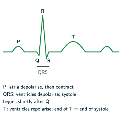
- An ECG is a graphical representation of the electrical activity of the heart during a cardiac cycle, obtained with an electro-cardiograph (NCERT page 200).
- NCERT illustrates connections at both wrists and the left ankle, calling them three leads (page 201). Distinguish the skin electrodes from a lead, an electrical view calculated from electrode signals. A standard clinical 12-lead ECG uses 10 electrodes: four limb and six chest electrodes (NHS Health Research Authority, “WEAR-TECH ECG”; NICE, KardiaMobile 6L final scope).
| Wave | What it shows |
|---|---|
| P-wave | atria depolarise, then contract |
| QRS complex | ventricles depolarise; systole begins shortly after Q |
| T-wave | ventricles repolarise; near the end of systole |
NCERT associates the T-wave’s end with the end of systole (page 201); electrical recovery and mechanical relaxation are related events, not identical measurements. Counting the QRS complexes in a given time gives the heart rate, and since ECGs from different people have roughly the same shape for a given lead configuration, a deviation indicates a possible abnormality or disease (page 201).
Solved examples
Example 1: Cardiac output
Using NCERT’s values, a stroke volume of 70 mL and a heart rate of 72 beats a minute, find the cardiac output.
Answer: 70 × 72 = 5040 mL a minute, about 5 litres, NCERT’s average of 5000 mL (page 200).
Example 2: Length of a cardiac cycle
How long is one cardiac cycle at 72 beats a minute? At 90?
Answer: 60 ÷ 72 = 0.83 s (NCERT gives 0.8 s, page 200); 60 ÷ 90 = 0.67 s.
Example 3: Heart rate from an ECG
An ECG strip shows 18 QRS complexes in 15 seconds. What is the heart rate?
Answer: each QRS complex represents one ventricular depolarisation; in this normal tracing, 18 beats in 15 s = 18 × 4 = 72 beats a minute.
Example 4: The pacemaker (NCERT Exercises 10 and 11)
Why is the sino-atrial node called the pacemaker, and what is the significance of the AVN and the AV bundle?
Answer: the SAN generates the maximum number of action potentials, 70–75 a minute, so it starts and maintains the rhythmic contraction of the heart. The action potential from the SAN reaches the ventricles through the AVN and the AV bundle, which runs through the septa and branches into Purkinje fibres in the ventricular muscle, so the ventricles contract after the atria (NCERT page 199).
Example 5: Heart sounds (NCERT Exercise 13)
Explain heart sounds.
Answer: two prominent sounds are heard in each cardiac cycle through a stethoscope: the first (lub), from the closure of the tricuspid and bicuspid valves at the start of ventricular systole, and the second (dub), from the closure of the semilunar valves as the ventricles relax. They are of clinical diagnostic significance (NCERT pages 199–200).
Example 6: Systole and diastole; P- and T-waves (NCERT Exercise 7c, d)
Write the differences between (c) systole and diastole, (d) the P-wave and T-wave.
Answer: (c) systole is the contraction of the atria or ventricles, which pushes blood onward; diastole is their relaxation, when they fill (NCERT pages 199–200). (d) The P-wave shows the depolarisation of the atria, which leads to their contraction; the T-wave shows the repolarisation of the ventricles, and NCERT associates its end with the end of systole (page 201).
Example 7: Evolution of the heart (NCERT Exercise 8)
Describe the evolutionary change in the pattern of the heart among the vertebrates.
Answer: fishes have a 2-chambered heart with single circulation; amphibians and most reptiles a 3-chambered heart, where oxygenated and deoxygenated blood mix in one ventricle (incomplete double circulation); crocodiles, birds and mammals a 4-chambered heart, and in birds and mammals the two kinds of blood are pumped out without mixing (double circulation) (NCERT page 197).
Common mistakes to avoid
Common mistake
Matching the heart sounds to the wrong valves
Lub is the closure of the tricuspid and bicuspid (atrio-ventricular) valves; dub is the closure of the semilunar valves (NCERT page 200).
Common mistake
Putting the SAN in the left atrium
Both the SAN and the AVN are in the right atrium: the SAN in its right upper corner, the AVN in its lower left corner (NCERT page 199).
Common mistake
Counting reptiles as 4-chambered
Reptiles have a 3-chambered heart, except crocodiles, which have 4 chambers (NCERT page 197).
Common mistake
Saying the T-wave starts systole
Ventricular contraction follows depolarisation (QRS). The T-wave records electrical recovery; NCERT associates its end with the end of systole (page 201).
Common mistake
Assuming every mollusc has an open system
NCERT gives arthropods and molluscs as open, and annelids and chordates as closed (page 197). Beyond the NCERT text, cephalopod molluscs such as octopuses and squid are an exception: they have a closed circulatory system (Villanueva and colleagues, Frontiers in Physiology, 2017). Another open system outside this list is that of the hemichordate Balanoglossus (NCERT Chapter 4, Section 4.2.10, page 45).
Common mistake
Doubling the cardiac output
Cardiac output is the volume pumped out by each ventricle per minute, about 5000 mL from each; do not add the two ventricles together (NCERT page 200).
Quick revision summary
- Open circulation (NCERT: arthropods and molluscs; beyond NCERT, cephalopods are closed) uses sinuses; closed circulation (annelids, chordates) keeps blood in vessels and can be regulated more precisely.
- Fishes: 2 chambers, single circulation; amphibians and reptiles (not crocodiles): 3 chambers, mixed blood; crocodiles, birds, mammals: 4 chambers; birds and mammals: double circulation.
- The human heart: pericardium; two atria, two ventricles; septa; tricuspid, bicuspid and semilunar valves for one-way flow; ventricles thicker-walled.
- Nodal tissue: SAN (pacemaker, 70–75 a minute), AVN, AV bundle, Purkinje fibres; the heart is autoexcitable.
- Cardiac cycle about 0.8 s; stroke volume about 70 mL; cardiac output about 5000 mL a minute; lub (AV valves) and dub (semilunar valves); ECG: P, QRS, T.
Formula sheet
- cardiac output = stroke volume × heart rate = 70 mL × 72 ≈ 5000 mL a minute
- cardiac cycle = 60 ÷ 72 ≈ 0.8 s
- lub: tricuspid and bicuspid close; dub: semilunar valves close
- P: atrial depolarisation; QRS: ventricular depolarisation; T: ventricular repolarisation
- SAN → AVN → AV bundle → right and left bundles → Purkinje fibres
Questions students ask
Why is the wall of the left ventricle thicker than that of the right ventricle?
The left ventricle has the thicker wall because it pumps blood through the aorta to the whole body, while the right ventricle sends blood only to the lungs (NCERT page 201); beyond the NCERT text, the lung circuit runs at a much lower pressure (Woulfe and Walker, Frontiers in Physiology, 2021). NCERT compares only the ventricles with the atria, whose walls are much thinner (page 199); each chamber’s wall thickness reflects the force it must generate (NCI SEER Training Modules, “Structure of the Heart”). At birth the two ventricle walls are about equally thick, as before birth the right ventricle also pumps blood to the body (Woulfe and Walker, as above).
What is the difference between the SA node and the AV node?
The SA node starts each heartbeat and the AV node passes it on to the ventricles: the SAN, in the right upper corner of the right atrium, generates the most action potentials, 70–75 a minute, and so is the pacemaker, while the AVN, in the lower left corner of the right atrium, hands the impulse to the AV bundle (NCERT page 199). Beyond the NCERT text, the AVN conducts slowly, giving the atrial contraction time to fill the ventricles, and it can take over as a backup pacemaker if the SA node fails (Park and Fishman, Journal of Cardiovascular Development and Disease, 2017).
Is one heartbeat the same as one cardiac cycle?
Yes, one heartbeat is one cardiac cycle: NCERT says the heart beats 72 times a minute, that is, it performs that many cardiac cycles a minute (page 200). Each cycle produces the two heart sounds, lub and dub, that can be heard through a stethoscope (page 200). So the length of one cycle is 60 seconds divided by the heart rate: at 60 beats a minute a cycle lasts 1 s, and at 120 beats a minute only 0.5 s.
Why does an ECG show no separate wave for atrial repolarisation?
An ECG shows no separate wave for atrial repolarisation because the atria repolarise at about the same time as the ventricles depolarise, so the small atrial signal is hidden by the large QRS complex (beyond the NCERT text; Ayano and colleagues, Diagnostics, 2022; Zyout and colleagues, Diagnostics, 2023). That is why a standard ECG has a P-wave for the atria but a T-wave only for the ventricles: NCERT’s T-wave is the return of the ventricles from the excited to the normal state (page 201).
What is the difference between an ECG, an EKG and an echocardiogram?
An ECG and an EKG are the same test, a recording of the heart’s electrical activity, while an echocardiogram is a different test that uses ultrasound to show the heart itself. NCERT defines the ECG as a graphical representation of the electrical activity of the heart during a cardiac cycle (page 200), and EKG is simply another abbreviation for the electrocardiogram (NHLBI, “Arrhythmias: Diagnosis”). Beyond the NCERT text, echocardiography is an ultrasound recording of the size and motion of the heart and its surrounding tissues, so it shows how the chambers and valves move rather than the electrical waves (MeSH, US National Library of Medicine, “Echocardiography”).
Practice questions
Try each question first, then open the answer to check your working.
-
Q1. Which valve guards the opening between the left atrium and the left ventricle?
Show answerHide answer
The bicuspid (mitral) valve (NCERT page 198).
-
Q2. By how much does atrial systole increase the flow of blood into the ventricles?
Show answerHide answer
By about 30 per cent (NCERT page 199).
-
Q3. Which three attachment sites does NCERT describe for its ECG illustration?
Show answerHide answer
Both wrists and the left ankle (NCERT page 201). These are attachment sites; a standard clinical 12-lead ECG uses 10 electrodes.